Review
. 2012 Apr;35(4):220-9.
doi: 10.1016/j.tins.2011.10.007. Epub 2011 Dec 15.

Sensing sound: molecules that orchestrate mechanotransduction by hair cells

Affiliations
Review

Sensing sound: molecules that orchestrate mechanotransduction by hair cells

Piotr Kazmierczak et al. Trends Neurosci. 2012 Apr.

Abstract

Animals use acoustic signals to communicate and to obtain information about their environment. The processing of acoustic signals is initiated at auditory sense organs, where mechanosensory hair cells convert sound-induced vibrations into electrical signals. Although the biophysical principles underlying the mechanotransduction process in hair cells have been characterized in much detail over the past 30 years, the molecular building-blocks of the mechanotransduction machinery have proved to be difficult to determine. We review here recent studies that have both identified some of these molecules and established the mechanisms by which they regulate the activity of the still-elusive mechanotransduction channel.


Figures

Fig. 1
Fig. 1. The mammalian auditory sense organ and its hair cells
(A) Diagram of the inner ear. The snail-shaped cochlea (end organ for the perception of sound) and parts of the vestibule (end organ for the perception of head movement) are indicated (panel modified from [4]). (B) Diagram of the organ of Corti. One inner hair cell (IHC) and three outer hair cells (OHCs) are indicated. (C) Scanning electron micrograph of the cochlear sensory epithelium of the mouse after removal of the tectorial membrane (kindly provided by Dr. Nicolas Grillet, TSRI). The image shows the stereociliary bundles of two IHCs. Note the staircase arrangement of the rows of stereocilia. Scale bar: 2 μm.
Fig. 2
Fig. 2. Hair cells and their mechanotransduction machinery
(A) Cross section through the apical part of a hair cell. Hair bundles consist of several rows of actin-rich stereocilia and a microtubule-based kinocilium. The sterocilia are connected to each other and to the kinocilium by extracellular filaments that can be visualized by electron microscopy. These are the tip links, top connectors, ankle links and kinociliary links (for a recent review see [4]). Note that the kinocilium, kinociliary links, ankle links and top connectors are present in murine cochlear hair cells only during hair bundle development. These structures degenerate once hair bundles have reached their mature shape and only stereocilia, tip links and top connectors remain [114]. (B) Diagram of the tip-link region, indicating molecules that are part of the tip-link complex. CDH23 homodimers form the upper part of the tip link and PCDH15 homodimers the lower part [59]. Two electron dense regions (shaded in gray) can be visualized by transmission electron microscopy in proximity to the upper and lower insertion sites of tip links [103]. Immunolocalization studies have localized the indicated proteins to the electron dense regions (for a recent review see [4]). (C) Current model of activation and adaptation of transduction channels in hair cells. Transduction channels that are located in proximity to the lower insertion site of tip links are opened by deflection of the hair bundle in the direction of the longest sterocilia. The tip link is thought to gate the channel. Ca2+ that flows into the stereocilia leads to fast adaptation likely by binding to the channel or a molecule nearby. Slow adaptation is thought to be regulated by a myosin motor complex at the upper insertion site of tip links. Upon Ca2+ entry, the adaptation motor is released from the cytoskeleton and slips down the actin filaments, leading to channel closure. Tension in the transduction complex is restored by movement of the myosin motor towards the tips of stereocilia (for a recent review see [1]). However, the localization of the transduction channel raises questions regarding models of slow adaptation because it places the site of Ca2+ entry into stereocilia at the lower tip-link end, far away from the proposed localization of the adaptation motor at the upper tip-link end. It has been proposed that Ca2+ entering through the transduction channel might affect the adaptation motor hooked up to the next tip link lower down in the same stereocilium [1], but adaptation motors in the longest stereocilia would then likely not show Ca2+-dependent adaptation.
Fig. 3
Fig. 3. Molecules of transduction
The domain structure of proteins that have been implicated as components of the tip-link complex are indicated and can be grouped into three categories: 1) transmembrane receptors of the cadherin superfamily (CDH23 and PCDH15) that form tip-link filaments [57-59]. The classical C-cadherin is shown for comparison to the tip-link cadherins. Note that the extracellular domains of CDH23 and PCDH15 are substantially larger than those of classical cadherins. The figure also shows different isoforms of CDH23 and PCDH15 that differ in their cytoplasmic domains. Two CDH23 isoforms have been identified in hair cells that are generated by alternative splicing of exon 68 [57, 86]. For PCDH15, three prominent isoforms that are generated by alternative splicing (PCDH15-CD1, -CD2, and -CD3) are expressed in hair cells [58, 87]. All CDH23 and PCDH15 isoforms contain consensus-binding sites for PDZ domain proteins (PBIs) (for a recent review see [61]). Which isoform of CDH23 and PCDH15 is at tip links remains to be determined; 2) adaptor proteins harmonin and sans. These adaptor proteins bind to the cytoplasmic domains of tip-link cadherins [86, 95-98, 115]. Harmonin can bind to the CDH23 and PCDH15 cytoplasmic domains but it co-localizes at tip links only with CDH23 [29]. Sans can bind to CDH23 and has been localized to the upper and lower ends of tip [91, 111]; 3) myosin motor proteins that are implicated in slow adaptation. Myo1c and Myo7a have been localized to the upper end of tip links but have also been reported, depending on species and experimental conditions, to be more broadly distributed in hair cells [105, 106, 108, 111, 116]. Abbreviations are as follows: Ank, ankyrin-like repeat; cc, coiled-coil domain; CEN, central sans domain; EC, extracellular cadherin repeat; FERM, protein 4.1, ezrin, radixin, moesin domain; IQ, calmodulin binding IQ domain; MyTH4, myosin tail homology 4 domain; N-ter, N-terminal domain of harmonin; PBI, PDZ binding interface; PDZ, PSD95/SAP90, Discs large, zonula occludens-1 domain; PST, proline, serine, threonine rich domain; SAM, sterile alpha motif domain; SH3, src homology 3 domain.
Figure 4
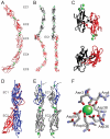
Figure 4. Structural features of the extracellular domains of classical cadherins and Cdh23
The figures summarize crystallographic data for classical cadherins and tip-link cadherins. (A) The entire Xenopus laevis C-cadherin extracellular domain (red) in the Ca2+ (green) bound state is shown. Note the five extracellular cadherin repeats (EC1-5), the three Ca2+ ions that are bound between each of the EC domains, and the curvature of the ectodomain [67]. (B) Adhesive trans dimer of murine E-cadherin [117]. Individual E-cadherin extracellular domains (red and black) emanate from opposing directions. The trans binding surface is located in the EC1 domain. Side view (left) reveals the characteristic curvature of the monomers, which appear straight when rotated by 90° (right). (C) The N-terminal EC1 domains of classical cadherins that engage in trans-interactions are depicted. The molecules form the so-called strand-swapped dimers. Classical type I cadherins such as murine E-cadherin (upper) contain in EC1 one key tryptophan residue (W2) that fits into a binding pocket in EC1 of its binding partner [117]. Classical type II cadherins such as murine MN-cadherin (lower) contain two tryptophan residues (W2 and W4) that fit into two binding pockets on EC1 of the binding partner [70]. (D) Crystallographic structure of the EC1-EC2 domain of murine CDH23 (blue) [71, 72]. The classic cadherin fold with Ca2+ ions (blue dots) between EC1 and EC2 is maintained. Superimposition of EC1-EC2 of C-cadherin (red) highlights the straight alignment of CDH23 EC1-EC2 compared to the curvature of C-cadherin [67, 71]. (E) Structure of the EC1-EC2 domains of two parallel CDH23 molecules. Note that only monomeric structures have been crystallized. The contact surface between EC domains that are aligned in parallel remain to be determined. Ca2+ ions (green dots) are indicated. Note the presence of a Ca2+ ion at the extreme N-terminus of CDH23 that is not bound by classical cadherins. The strands formed by amino acids 1-4 are clamped down in a loop (blue) by the coordinate bond between Asn3, Arg4, Asp36, Asp38, Asp40 and Ca2+ to form a large exposed area on the surface of EC1. Mutations in Asn3 and Arg4 reduce CDH23 binding to PCDH15 [71]. (F) Detailed view of the amino acids in CDH23 that coordinate the N-terminal Ca2+ [71, 72]. Structures were generated in RasMol (http://www.umass.edu/microbio/rasmol/) using protein coordinates from the Protein Data Bank (PDB) database (www.pdb.org).
Figure 5
Figure 5. Structural aspects of the UTLD complex emerging from crystallographic and biochemical studies
(A,B) Model of the interactions between proteins at the UTLD. In (A) a model of the interaction surfaces is provided that is based on biochemical data and, where available, on structural information [86, 95-102]. In (B), the known crystal structures are shown, except for an internal binding motif for PDZ-domain proteins (NBI) of CDH23, which was fitted artificially into its N-terminal binding site on harmonin. Protein-domains in (A) are color-coded to match the structures in (B). Abbreviations are as in Figure 3. Note that we present here a partial representation not considering all interactions revealed by biochemical data. For example, Myo7a can also bind to harmonin and CDH23 [94, 106], but these complexes have not yet been crystallized. Structures were generated as described in Figure 4.

Similar articles

Cited by

References

    1. Gillespie PG, Muller U. Mechanotransduction by hair cells: models, molecules, and mechanisms. Cell. 2009;139:33–44. - PMC - PubMed
    1. Meyer AC, Moser T. Structure and function of cochlear afferent innervation. Curr Opin Otolaryngol Head Neck Surg. 2010;18:441–446. - PubMed
    1. Richardson GP, et al. How the genetics of deafness illuminates auditory physiology. Annual review of physiology. 2011;73:311–334. - PubMed
    1. Schwander M, et al. Review series: The cell biology of hearing. J Cell Biol. 2010;190:9–20. - PMC - PubMed
    1. Pickles JO, et al. Cross-links between stereocilia in the guinea pig organ of Corti, and their possible relation to sensory transduction. Hear Res. 1984;15:103–112. - PubMed

Publication types