. 2012 Jan 10;22(1):12-20.
doi: 10.1016/j.cub.2011.11.028. Epub 2011 Dec 15.

Genetic dissection reveals two separate retinal substrates for polarization vision in Drosophila

Affiliations

Genetic dissection reveals two separate retinal substrates for polarization vision in Drosophila

Mathias F Wernet et al. Curr Biol. .

Abstract

Background: Linearly polarized light originates from atmospheric scattering or surface reflections and is perceived by insects, spiders, cephalopods, crustaceans, and some vertebrates. Thus, the neural basis underlying how this fundamental quality of light is detected is of broad interest. Morphologically unique, polarization-sensitive ommatidia exist in the dorsal periphery of many insect retinas, forming the dorsal rim area (DRA). However, much less is known about the retinal substrates of behavioral responses to polarized reflections.

Summary: Drosophila exhibits polarotactic behavior, spontaneously aligning with the e-vector of linearly polarized light, when stimuli are presented either dorsally or ventrally. By combining behavioral experiments with genetic dissection and ultrastructural analyses, we show that distinct photoreceptors mediate the two behaviors: inner photoreceptors R7+R8 of DRA ommatidia are necessary and sufficient for dorsal polarotaxis, whereas ventral responses are mediated by combinations of outer and inner photoreceptors, both of which manifest previously unknown features that render them polarization sensitive.

Conclusions: Drosophila uses separate retinal pathways for the detection of linearly polarized light emanating from the sky or from shiny surfaces. This work establishes a behavioral paradigm that will enable genetic dissection of the circuits underlying polarization vision.


Figures

Figure 1
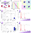
Figure 1. Drosophila manifests orientation responses to linearly polarized stimuli presented either dorsally, or ventrally
A. Schematic of the experimental setup used to present linearly polarized UV light from above to populations of Drosophila, which were filmed in the infrared from below. A polarization filter (HN42HE) was facing the flies, with 2 sheets of diffuser paper facing the light source. IR = infrared light. UV POL = polarized UV light. Diff = Diffuser. Pol = Polarizer. B. Summary of the stimulus protocol used. A computer-controlled servomotor rotated the polarization filter in 45° increments, remaining still for 5 seconds at each position. Different motor positions are denoted with different colors. C. Polar histograms of fly angular headings are shown for dorsally stimulated flies at each motor position. D. Basic description of wild type polarotactic responses for linearly polarized UV stimulus presented dorsally. White bars symbolize UV-POL stimulation, green and blue bars stimulation with polarized light of the respective color (see methods). All error bars = +/− 1 S.E.M. *=p<0.05, **p<0.01, ***p<0.001. n.s. = not significant. E. Alignment values A, plotted as a function of dorsal UV stimulus intensity. Dashed line: intensity setting used for all subsequent experiments. Red box: UV intensity of skylight at dusk (Palo Alto, CA – see Supplemental Experimental Procedures). F. Basic description of wild type responses for linearly polarized UV stimulus presented ventrally. G. A values plotted as a function of ventral UV stimulus intensity.
Figure 2
Figure 2. Dorsal polarotactic behavior is mediated by the ‘Dorsal Rim Area’
A. Testing the necessity of photoreceptor subtypes mediating behavioral responses to UV-POL stimuli presented dorsally. Polarotactic responses were measured in flies expressing UAS-shibirets under the control of GAL4 drivers expressed in various subtypes of photoreceptors. Unlabeled bars: not significantly different from the control. B. Sufficiency of photoreceptor subtypes mediating behavioral responses to POL stimuli presented dorsally. Opsin drivers (both wild type and mutated) and hth-GAL4 were used to rescue photoreceptor function by expressing eye-specific Phospholipase C (NorpA) from newly generated UAS-norpA transgenes (shown schematically, see methods), in norpA/norpA mutant flies. Open bars denote experimental genotypes, gray bars denote negative controls (a norpA/norpA mutant, bearing UAS-norpA, without a GAL4 driver).
Figure 3
Figure 3. Rhabdomeres of inner photoreceptors in the DRA are untwisted
Ommadidia of the Dorsal Rim Area (DRA) and of the adjacent dorsal area (DA) were studied A, D Transmission electron micrographs showing rhabdom structure (a) and microvilli orientation of R7 in individual ommatidia at distal (b) and proximal (c) levels of R7. Numbers indicate receptor types. Straight lines in rhabdomere cross sections give microvilli orientations. Calibration bars 1 μm. B, C Total range of microvilli directions expressed by fans in different groups of ommatidia (same as in line graphs E, F). Red fans represent R7, blue fans R8. Fine black line marks the boundary between the DRA and the DA. Fat black line shows the eye rim. Interrupted arrowed line is the v-axis of the ommatidial pattern pointing dorsal (compare Figure S3D). dco: dorso-caudal origin of ommatidial rows. Calibration bars 10 μm. Note that one ommatidium has a R7DRA (large, non-twisting rhabdomere) but a R8DA (small, twisting rhabdomere). E–F Graphic representation of microvilli orientation at different retinal levels (twist functions) in R7 (left family of curves) and R8 (right family of curves). The ordinate indicates microvilli orientation relative to a straight line through the centers of R1 and R3 rhabdomeres (0°; stipled line). The abscissa gives retinal level relative to the surface of the eye (0 μm indicates level of first section containing rhabdoms). Colors mark data from different, identified ommatidia.
Figure 4
Figure 4. Ventral R7 photoreceptors can mediate polarotactic responses
A. Behavioral responses to a ventral POL stimulus after inactivation of photoreceptor subtypes with two copies of UAS-shibirets. Open bars denote experimental genotypes, gray bars denote control genotypes. B. No single photoreceptor subtype is required for an orientation response of upside-down walking flies to linearly polarized green light, but behavior gets strongly abrogated upon simultaneous inactivation of rh1- and [rh5+rh6] subtypes, and completely disappears using 3 copies of UAS-shibirets (compare dark and light green bars).
Figure 5
Figure 5. Moderate- and low-twist R7 cells exist in the ventral eye
Three different groups of ommatidia in the ventral eye (VA1, VA2, VA3). Rhabdomeres generally twist but the amount of twist and the shape of the twist functions differed between groups. A, D as in Figure 2 A, D. Calibration bars 1 μm. B, C, E, F as in Figure 2 B, C (same ommatidia as in line graphs G,H). White asterisks on some fans in C indicate that data for the most proximal rhabdomere are missing. vfo ventro-frontal, vco ventro-caudal origin of ommatidial rows. Calibration bars 10 μm. G–I as in Figure 2 E, F.
Figure 6
Figure 6. Outer photoreceptors R1–R6 contribute to ventral polarotaxis
A. NorpA rescue experiments (open bars, and shaded bar control) were used to define photoreceptor sub-type sufficiency for behavioral responses to polarized UV light. B. Orienting responses to polarized green light in norpA rescue experiments. Alignment responses upon outer photoreceptors rescue (rh1-norpA) were eliminated by the QWP, and restored by rotating it 45°. C. Twist functions of receptors R1, R2, R4 and R5. Typical twist functions of each cell type in three ommatidia are shown. The twist functions of R4 and R5 are generally flatter than those of R1 and R2. D. Polarization sensitivity (PS) of receptor types R1–8. PS of samples of 4, 5 and 8 ommatidia in three different eyes are shown. Black circles indicate average PS. Note that R4, R5 and R6 have higher PS than R1–3. E. Model summarizing photoreceptor contributions to linearly polarized stimuli presented to the dorsal, or the ventral retina, respectively. Left: insects encounter linearly polarized light originating from atmospheric scattering, or from reflections off of shiny surfaces such as water. Middle: schematic representation of the dorsal half (top) or ventral half (bottom) of the fly retina (necessary ommatidia are labeled red), followed by a schematic representation of photoreceptor classes in these ommatidia (photoreceptors that provide input to UV polarotaxis, green polarotaxis, or both behaviors are shown in violet, green, and blue, respectively). Right: photoreceptor types providing behavioral contributions. Behavioral output is symbolized by a sinusoid function, synergistic interactions between photoreceptor subtypes are symbolized by a ‘+’ sign.

Comment in

References

    1. Wehner R. Polarization vision – a uniform sensory capacity? J Exp Biol. 2001;204:2589–2596. - PubMed
    1. Wehner R, Labhart T. Polarization vision. In: Warrant EJ, Nilsson D-E, editors. Invertebrate vision. Cambridge: 2006.
    1. Nilsson DE, Warrant EJ. Visual discrimination: Seeing the third quality of light. Curr Biol. 1999;9:R535–537. - PubMed
    1. Rossel S. Navigation by bees using polarized skylight. Comp Biochem Physiol. 1993;104A:695–708.
    1. Wehner R. Desert ant navigation: how minibrains solve complex tasks. J Comp Physiol A. 2003;189:579–588. - PubMed

Publication types

LinkOut - more resources