Ictal MEG onset source localization compared to intracranial EEG and outcome: improved epilepsy presurgical evaluation in pediatrics
- PMID: 22178034
- PMCID: PMC3520066
- DOI: 10.1016/j.eplepsyres.2011.11.007
Ictal MEG onset source localization compared to intracranial EEG and outcome: improved epilepsy presurgical evaluation in pediatrics
Abstract
Purpose: Magnetoencephalography (MEG) has been shown a useful diagnostic tool for presurgical evaluation of pediatric medically intractable partial epilepsy as MEG source localization has been shown to improve the likelihood of seizure onset zone (SOZ) sampling during subsequent evaluation with intracranial EEG (ICEEG). We investigated whether ictal MEG onset source localization further improves results of interictal MEG in defining the SOZ.
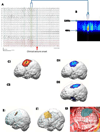
Methods: We identified 20 pediatric patients with one habitual seizure during MEG recordings between October 2007 and April 2011. MEG was recorded with sampling rates of 600Hz and 4000Hz for 10 and 2min respectively. Continuous head localization (CHL) was applied. Source localization analyses were applied using multiple algorithms, both at the beginning of ictal onset and for interictal MEG discharges. Ictal MEG onsets were identified by visual inspection and power spectrum using short-time Fourier transform (STFT). Source localizations were compared with ICEEG, surgical procedure and outcome.
Key findings: Eight patients met all inclusion criteria. Five of the 8 patients (63%) had concordant ictal MEG onset source localization and interictal MEG discharge source localizations in the same lobe, but the source of ictal MEG onset was closer to the SOZ defined by ICEEG.
Significance: Although the capture of seizures during MEG recording is challenging, the source localization for ictal MEG onset proved to be a useful tool for presurgical evaluation in our pediatric population with medically intractable epilepsy.
Copyright © 2011 Elsevier B.V. All rights reserved.
Figures


Similar articles
-
Interictal and ictal source localization for epilepsy surgery using high-density EEG with MEG: a prospective long-term study.Brain. 2019 Apr 1;142(4):932-951. doi: 10.1093/brain/awz015. Brain. 2019. PMID: 30805596 Free PMC article.
-
Accuracy of MEG in localizing irritative zone and seizure onset zone: Quantitative comparison between MEG and intracranial EEG.Epilepsy Res. 2016 Nov;127:291-301. doi: 10.1016/j.eplepsyres.2016.08.013. Epub 2016 Aug 16. Epilepsy Res. 2016. PMID: 27693985
-
Ictal magnetoencephalography in temporal and extratemporal lobe epilepsy.Epilepsia. 2003 Oct;44(10):1320-7. doi: 10.1046/j.1528-1157.2003.14303.x. Epilepsia. 2003. PMID: 14510826
-
Use of magnetoencephalography in the presurgical evaluation of epilepsy patients.Clin Neurophysiol. 2007 Jul;118(7):1438-48. doi: 10.1016/j.clinph.2007.03.002. Epub 2007 Apr 23. Clin Neurophysiol. 2007. PMID: 17452007 Review.
-
[High-resolution EEG (HR-EEG) and magnetoencephalography (MEG)].Neurochirurgie. 2008 May;54(3):185-90. doi: 10.1016/j.neuchi.2008.02.014. Epub 2008 Apr 15. Neurochirurgie. 2008. PMID: 18417162 Review. French.
Cited by
-
Source localization of the seizure onset zone from ictal EEG/MEG data.Hum Brain Mapp. 2016 Jul;37(7):2528-46. doi: 10.1002/hbm.23191. Epub 2016 Apr 5. Hum Brain Mapp. 2016. PMID: 27059157 Free PMC article.
-
The role of functional neuroimaging in pre-surgical epilepsy evaluation.Front Neurol. 2014 Mar 24;5:31. doi: 10.3389/fneur.2014.00031. eCollection 2014. Front Neurol. 2014. PMID: 24715886 Free PMC article. Review.
-
Lateralization Value of Low Frequency Band Beamformer Magnetoencephalography Source Imaging in Temporal Lobe Epilepsy.Front Neurol. 2018 Oct 5;9:829. doi: 10.3389/fneur.2018.00829. eCollection 2018. Front Neurol. 2018. PMID: 30344505 Free PMC article.
-
Source imaging of seizure onset predicts surgical outcome in pediatric epilepsy.Clin Neurophysiol. 2021 Jul;132(7):1622-1635. doi: 10.1016/j.clinph.2021.03.043. Epub 2021 Apr 28. Clin Neurophysiol. 2021. PMID: 34034087 Free PMC article.
-
Practical Fundamentals of Clinical MEG Interpretation in Epilepsy.Front Neurol. 2021 Oct 14;12:722986. doi: 10.3389/fneur.2021.722986. eCollection 2021. Front Neurol. 2021. PMID: 34721261 Free PMC article. Review.
References
-
- Assaf BA, Karkar KM, Laxer KD, Garcia PA, Austin EJ, Barbaro NM, Aminoff MJ. Ictal magnetoencephalography in temporal and extratemporal lobe epilepsy. Epilepsia. 2003;44:1320–1327. - PubMed
-
- Bollo RJ, Kalhorn SP, Carlson C, Haegeli V, Devinsky O, Weiner HL. Epilepsy surgery and tuberous sclerosis complex: special considerations. Neurosurg Focus. 2008;25:E13. - PubMed
-
- Brookes MJ, Stevensona CM, Barnesb GR, Hillebrandb A, Simpsonb MIG, Francisa ST, Morrisa PG. Beamformer reconstruction of correlated sources using a modified source model. NeuroImage. 2007;34:1454–1465. - PubMed
-
- Canuet L, Ishii R, Iwase M, Kurimoto R, Ikezawa K, Azechi M, Wataya-Kaneda M, Takeda M. Tuberous sclerosis: Localizing the epileptogenic tuber with synthetic aperture magnetometry with excess kurtosis analysis. J Clin Neurosci. 2008;15:1296–1298. - PubMed
Publication types
MeSH terms
Grants and funding
LinkOut - more resources
Full Text Sources
Medical
Miscellaneous

