

mTOR activates hypoxia-inducible factor-1α and inhibits neuronal apoptosis in the developing rat brain during the early phase after hypoxia-ischemia
- PMID: 22178140
- PMCID: PMC3525671
- DOI: 10.1016/j.neulet.2011.11.058
mTOR activates hypoxia-inducible factor-1α and inhibits neuronal apoptosis in the developing rat brain during the early phase after hypoxia-ischemia
Abstract
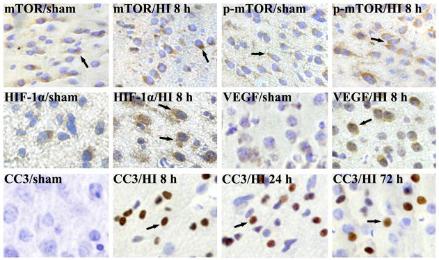
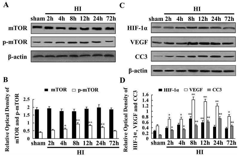
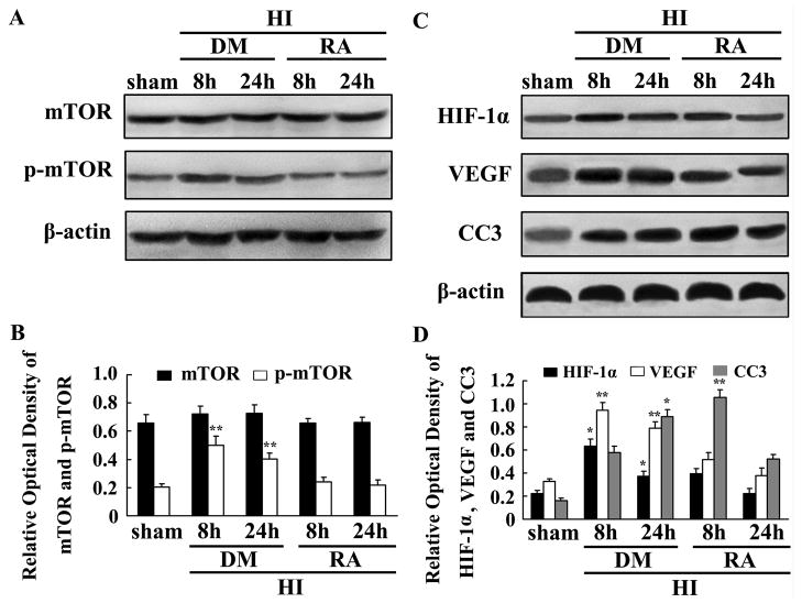
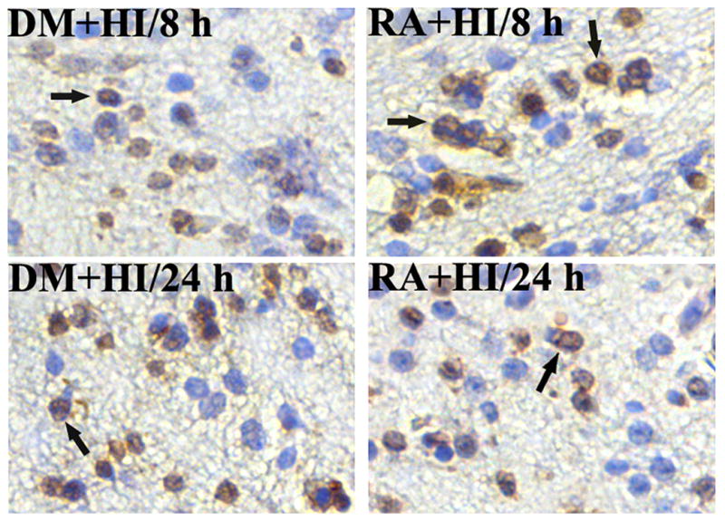
The mammalian target of rapamycin (mTOR) exerts neuroprotective effects under hypoxic or ischemic conditions. To explore whether mTOR participates in neuroprotective signaling through regulation of hypoxia-inducible factor-1α (HIF-1α), vascular endothelial growth factor (VEGF) and neuronal apoptosis in developing rat brain with hypoxia-ischemia (HI), we operated on postnatal day 10 rats by ligating the common carotid artery followed by exposure to systemic hypoxia. Brains were collected at various intervals to detect the expression of mTOR, phosphorylated mTOR (p-mTOR), HIF-1α, VEGF and cleaved caspase 3 (CC3), using immunohistochemistry and Western blot analysis. We also used terminal deoxynucleotidyl transferase-mediated dUTP-nick end labeling (TUNEL) to detect neuronal apoptosis. The p-mTOR protein expression increased at 2h after HI, peaked at 8h, lasted 24h, and then dropped to the basal level. Also, the expression of HIF-1α and VEGF was significantly enhanced and peaked at 8h after HI. Up-regulated expression of CC3 was observed at 2h, peaked at 24h, and lasted 72h after HI. Increased neuronal apoptosis is associated with reduced HIF-1α and VEGF expression. Furthermore, pretreatment with rapamycin, a mTOR specific inhibitor, significantly inhibited HIF-1α and VEGF protein after HI. The expression of CC3 and the number of TUNEL-positive cells were up-regulated at 8h and down-regulated at 24h after HI in the rapamycin-treated group. Our findings suggest that mTOR may participate in the regulation of HIF-1α, VEGF and neuronal apoptosis, serving neuroprotective functions after HI in developing rat brain.
Copyright © 2011 Elsevier Ireland Ltd. All rights reserved.
Conflict of interest statement
All of the authors involved in preparation of the above manuscript declare no conflict of interest in any form.
Figures
Similar articles
-
Relationship between HIF-1alpha expression and neuronal apoptosis in neonatal rats with hypoxia-ischemia brain injury.Brain Res. 2007 Nov 14;1180:133-9. doi: 10.1016/j.brainres.2007.08.059. Epub 2007 Sep 5. Brain Res. 2007. PMID: 17920049
-
[ROLE OF EXTRACELLULAR SIGNAL-RELATED PROTEIN KINASE 1/2 PATHWAY IN GINSENOSIDE Rg1 MEDIATED ANTI-APOPTOTIC EFFECT ON NEURON AFTER HYPOXIA ISCHEMIA BRAIN DAMAGE IN NEONATAL RATS].Zhongguo Xiu Fu Chong Jian Wai Ke Za Zhi. 2016 Aug 8;30(8):1011-1018. doi: 10.7507/1002-1892.20160204. Zhongguo Xiu Fu Chong Jian Wai Ke Za Zhi. 2016. PMID: 29786234 Chinese.
-
The involvement of phosphoinositid 3-kinase/Akt pathway in the activation of hypoxia-inducible factor-1alpha in the developing rat brain after hypoxia-ischemia.Brain Res. 2008 Mar 4;1197:152-8. doi: 10.1016/j.brainres.2007.12.059. Epub 2008 Jan 3. Brain Res. 2008. PMID: 18241842
-
The role and regulation of hypoxia-inducible factor-1alpha expression in brain development and neonatal hypoxic-ischemic brain injury.Brain Res Rev. 2009 Dec 11;62(1):99-108. doi: 10.1016/j.brainresrev.2009.09.006. Epub 2009 Sep 25. Brain Res Rev. 2009. PMID: 19786048 Review.
-
The role of mTOR signalling pathway in hypoxia-induced cognitive impairment.Int J Neurosci. 2021 May;131(5):482-488. doi: 10.1080/00207454.2020.1746308. Epub 2020 Apr 2. Int J Neurosci. 2021. PMID: 32202188 Review.
Cited by
-
Neuroprotective effects of DAHP and Triptolide in focal cerebral ischemia via apoptosis inhibition and PI3K/Akt/mTOR pathway activation.Front Neuroanat. 2015 Apr 22;9:48. doi: 10.3389/fnana.2015.00048. eCollection 2015. Front Neuroanat. 2015. PMID: 25954164 Free PMC article.
-
Neuroprotective Effects of Oxymatrine on PI3K/Akt/mTOR Pathway After Hypoxic-Ischemic Brain Damage in Neonatal Rats.Front Pharmacol. 2021 Apr 13;12:642415. doi: 10.3389/fphar.2021.642415. eCollection 2021. Front Pharmacol. 2021. PMID: 33927621 Free PMC article.
-
A preview of selected articles.Stem Cells Transl Med. 2021 Mar;10(3):333-336. doi: 10.1002/sctm.21-0036. Stem Cells Transl Med. 2021. PMID: 33619898 Free PMC article. No abstract available.
-
Mammalian target of rapamycin cell signaling pathway contributes to the protective effects of ischemic postconditioning against stroke.Stroke. 2014 Sep;45(9):2769-76. doi: 10.1161/STROKEAHA.114.005406. Epub 2014 Jul 10. Stroke. 2014. PMID: 25013017 Free PMC article.
-
Hypoxia inducible factor 1α promotes survival of mesenchymal stem cells under hypoxia.Am J Transl Res. 2017 Mar 15;9(3):1521-1529. eCollection 2017. Am J Transl Res. 2017. PMID: 28386377 Free PMC article.
References
-
- Balduini W, Carloni S, Buonocore G. Autophagy in hypoxia-ischemia induced brain injury: evidence and speculations. Autophagy. 2009;5(2):221–223. - PubMed
-
- Blomgren K, Leist M, Groc L. Pathological apoptosis in the developing brain. Apoptosis. 2007;12(5):993–1010. - PubMed
-
- Carloni S, Buonocore G, Balduini W. Protective role of autophagy in neonatal hypoxia-ischemia induced brain injury. Neurobiol Dis. 2008;32(3):329–339. - PubMed
-
- Del Bufalo D, Ciuffreda L, Trisciuoglio D, Desideri M, Cognetti F, Zupi G, Milella M. Antiangiogenic potential of the mammalian target of rapamycin inhibitor temsirolimus. Cancer Res. 2006;66(11):5549–5554. - PubMed
-
- Fahling M. Cellular oxygen sensing, signalling and how to survive translational arrest in hypoxia. Acta Physiol (Oxf) 2009;195(2):205–230. - PubMed
Publication types
MeSH terms
Substances
Grants and funding
LinkOut - more resources
Full Text Sources
Research Materials
Miscellaneous
