. 2012 Jan;233(1):534-42.
doi: 10.1016/j.expneurol.2011.11.039. Epub 2011 Dec 8.

Dysfunction of nodes of Ranvier: a mechanism for anti-ganglioside antibody-mediated neuropathies

Affiliations

Dysfunction of nodes of Ranvier: a mechanism for anti-ganglioside antibody-mediated neuropathies

Keiichiro Susuki et al. Exp Neurol. 2012 Jan.

Abstract

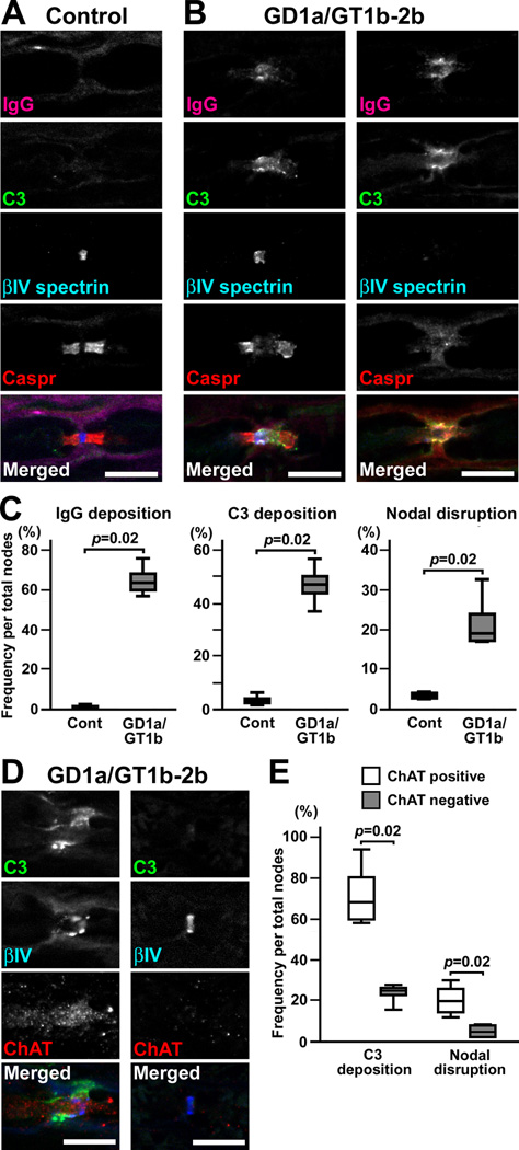
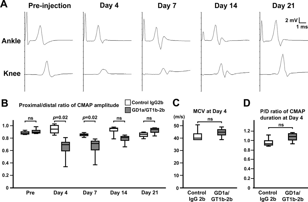
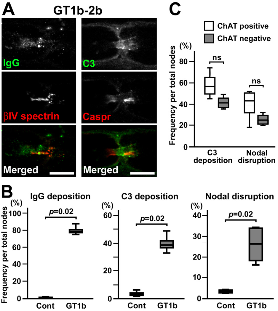
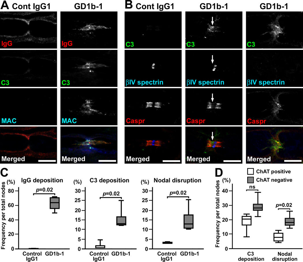
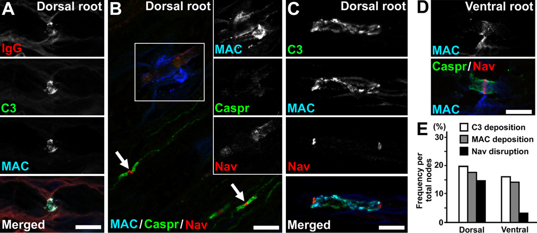
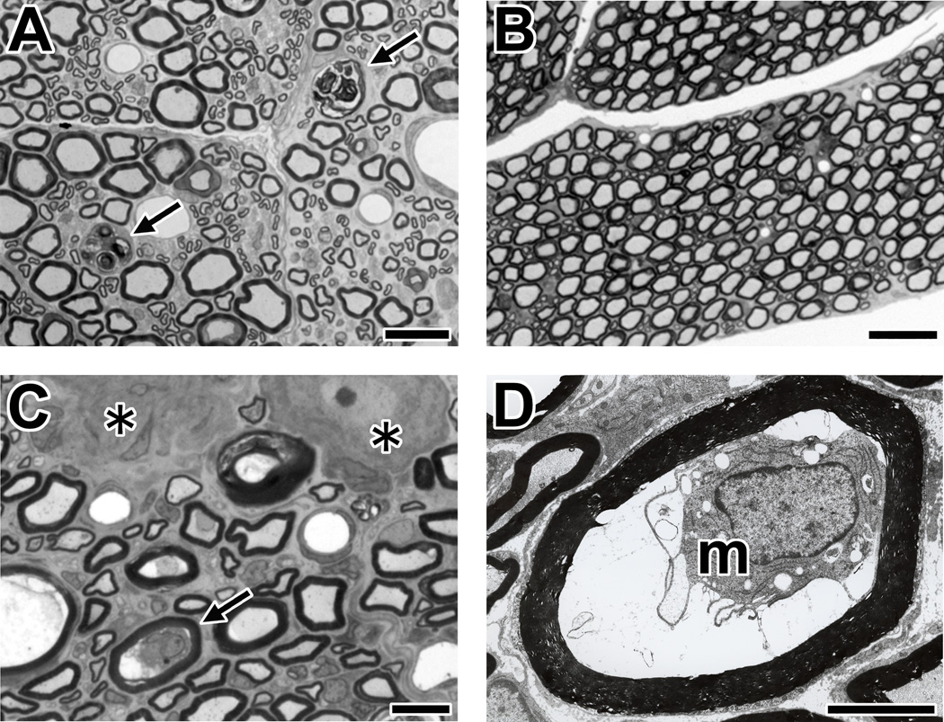
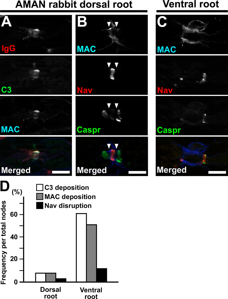
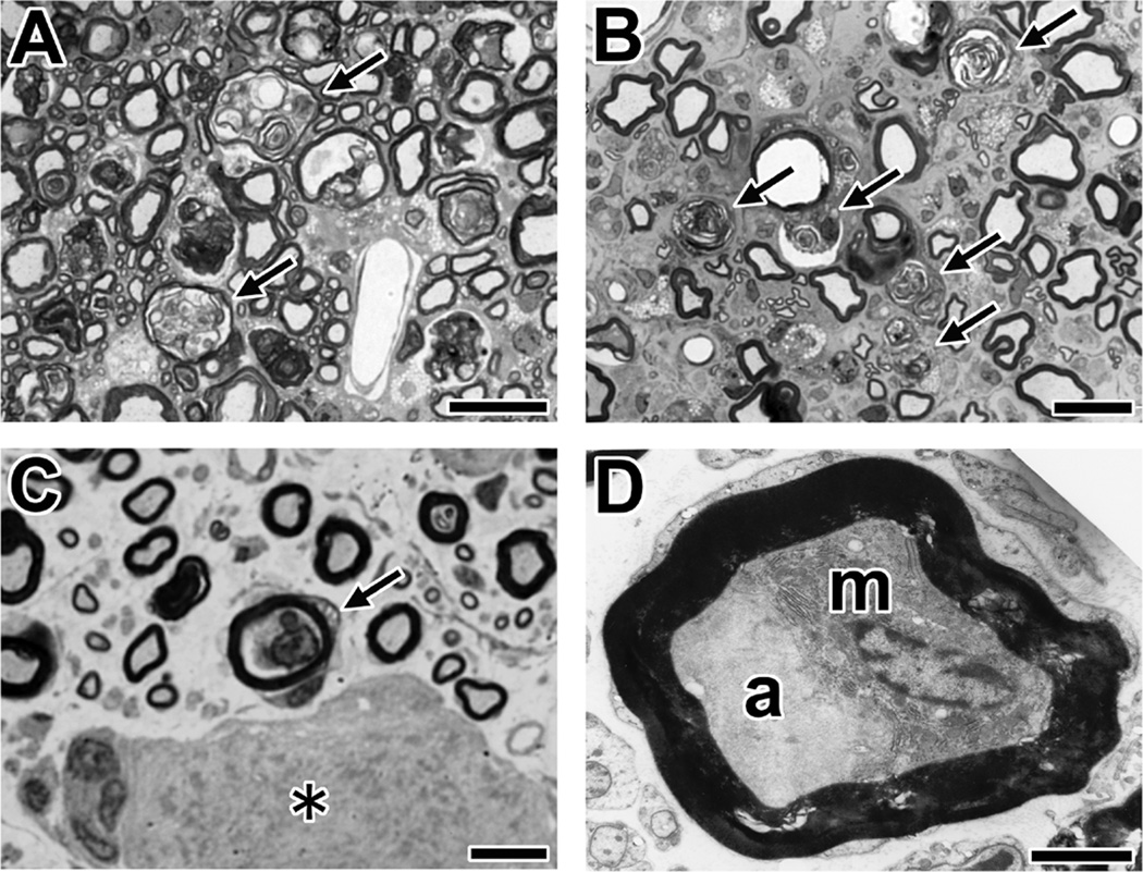
Autoantibodies against gangliosides GM1 or GD1a are associated with acute motor axonal neuropathy (AMAN) and acute motor-sensory axonal neuropathy (AMSAN), whereas antibodies to GD1b ganglioside are detected in acute sensory ataxic neuropathy (ASAN). These neuropathies have been proposed to be closely related and comprise a continuous spectrum, although the underlying mechanisms, especially for sensory nerve involvement, are still unclear. Antibodies to GM1 and GD1a have been proposed to disrupt the nodes of Ranvier in motor nerves via complement pathway. We hypothesized that the disruption of nodes of Ranvier is a common mechanism whereby various anti-ganglioside antibodies found in these neuropathies lead to nervous system dysfunction. Here, we show that the IgG monoclonal anti-GD1a/GT1b antibody injected into rat sciatic nerves caused deposition of IgG and complement products on the nodal axolemma and disrupted clusters of nodal and paranodal molecules predominantly in motor nerves, and induced early reversible motor nerve conduction block. Injection of IgG monoclonal anti-GD1b antibody induced nodal disruption predominantly in sensory nerves. In an ASAN rabbit model associated with IgG anti-GD1b antibodies, complement-mediated nodal disruption was observed predominantly in sensory nerves. In an AMAN rabbit model associated with IgG anti-GM1 antibodies, complement attack of nodes was found primarily in motor nerves, but occasionally in sensory nerves as well. Periaxonal macrophages and axonal degeneration were observed in dorsal roots from ASAN rabbits and AMAN rabbits. Thus, nodal disruption may be a common mechanism in immune-mediated neuropathies associated with autoantibodies to gangliosides GM1, GD1a, or GD1b, providing an explanation for the continuous spectrum of AMAN, AMSAN, and ASAN.


Conflict of interest statement

Conflict of interest: The authors declare no competing financial interests.

Figures

Fig. 1
Fig. 1. GD1a/GT1b-2b disrupts predominantly motor nerve nodes via the complement pathway
(A,B) Immunofluorescence analyses in longitudinal sections of rat sciatic nerves injected with control IgG2b or GD1a/GT1b-2b. Nerve fibers run horizontally in all panels. Sections are stained by antibodies to mouse IgG (magenta), C3 component of complement (green), βIV spectrin (blue), and Caspr (red). Control IgG2b weakly binds to the outer surface of myelin, but no C3 deposition is seen (A). Deposition of GD1a/GT1b-2b and C3 is associated with the abnormally lengthened gap between paranodal Caspr clusters with preserved nodal βIV spectrin (B, left column), or with completely damaged βIV spectrin and Caspr clusters (B, right column). (C) Frequency of immune-mediated nodal disruption after repeated injection of GD1a/GT1b-2b in rat sciatic nerves at day 4. The depositions of IgG or C3, or disrupted nodes are significantly more frequent by injection of GD1a/GT1b-2b compared to control IgG2b (n=4 in each group). For quantification, 200–300 nodes were observed in each rat sciatic nerve. +(D) Representative images of nodes in sciatic nerves at day 4 after repeated injection of GD1a/GT1b-2b together with complement. Left column shows disrupted node (shown by βIV spectrin in blue) with C3 deposition (green) in ChAT positive axon (red). Right column shows preserved node in ChAT negative axon. (E) Quantitation of C3 deposition and nodal disruption in ChAT positive or negative nodes at day 4. Data were collected from four animals. Scale bars = 10 µm (A,B,D).
Fig. 2
Fig. 2. Serial nerve conduction study in GD1a/GT1b-2b injected nerves
Control IgG2b or GD1a/GT1b-2b was injected into rat tibial nerves half way between ankle and knee twice (days 0 and 3). Nerve conduction study of tibial nerve was performed serially at different time points: pre-injection, days 4, 7, 14, and 21. (A) Representative wave forms from one animal injected with GD1a/GT1b-2b. At day 4, the amplitude after stimulation at the knee is abnormally reduced. Temporal dispersion is not apparent. The abnormal amplitude reduction disappears by day 21 without apparent late components. (B) Temporal changes of P/D ratio of CMAP amplitude (between the base line and negative peak) in rat tibial nerve injected with control IgG2b or GD1a/GT1b-2b. The same animals were analyzed for serial nerve conduction study (n=4 in each group). At days 4 and 7, P/D ratio of CMAP amplitude was significantly reduced in GD1a/GT1b-2b injected nerves. At day 14, the ratio still tended to be lower than control, but the difference did not reach statistical significance. By day 21, the ratio returned to normal. (C) There is no significant difference of MCV in the ankle-knee segment of tibial nerves between control and GD1a/GT1b-2b groups at day 4 (n=4 in each group). (D) There is no significant difference of P/D ratio of CMAP duration (from the onset to the final return to baseline) between control and GD1a/GT1b-2b groups at day 4 (n=4 in each group).
Fig. 3
Fig. 3. Nodal lesions caused by injection of GT1b-2b
(A) Immunofluorescent studies in longitudinal sections of rat sciatic nerves injected with GT1b-2b. Nerve fibers run horizontally in all panels. Left column shows deposition of mouse IgG (green) and a disrupted nodal βIV spectrin cluster (red). Right column shows deposition of C3 (green) and altered paranodal Caspr staining (red). (B) Frequency of immune-mediated nodal disruption by GT1b-2b. The depositions of IgG or C3, or disrupted nodes are significantly more frequent by injection of GT1b-2b compared to control IgG2b (n=4 in each group). Two hundred-300 nodes were observed in each nerve. (C) Quantitation of C3 deposition and nodal disruption in ChAT positive or negative nodes in GT1b-2b injected nerves. Data were collected from four animals. Scale bars = 10 µm (A).
Fig. 4
Fig. 4. Nodal lesions predominantly in sensory fibers caused by injection of GD1b-1
(A,B) Immunofluorescent studies in longitudinal sections of rat sciatic nerves injected with control IgG1 or GD1b-1. Nerve fibers run horizontally in all panels. (A) Sections are stained by antibodies to mouse IgG (red), C3 (green), and MAC (blue). Nonspecific binding of mouse control IgG1 is seen along the outer surface of the myelin sheath, but no staining on the axolemma was observed (left column). There are no apparent depositions of C3 or MAC. In GD1b-1 injected nerve (right column), depositions of IgG, C3, and MAC are seen on the axolemma at and near node. (B) Sections are stained by antibodies to C3 (green), βIV spectrin (blue), and Caspr (red). The nerve injected with control IgG1 does not show nodal C3 deposition, and clusters of nodal βIV spectrin and paranodal Caspr remain intact (left column). In the nerves injected with GD1b-1, the Caspr cluster on the left side seems to be reduced in association with C3 deposition, resulting in widening of the nodal gap (middle column, arrows). At the node with massive C3 deposition, both βIV spectrin and Caspr clusters are remarkably disrupted or disappear (right column). (C) Frequency of immune-mediated nodal disruption by GD1b-1. The depositions of IgG or C3, or disrupted nodes are significantly more frequent by injection of GD1b-1 compared to control IgG1 (n=4 in each group). Two hundred-300 nodes were observed in each nerve. (D) Quantitation of C3 deposition and nodal disruption in ChAT positive or negative nodes in GD1b-1 injected nerves. Data were collected from four animals. Scale bars = 10 µm (A,B).
Fig. 5
Fig. 5. Disruption of sensory nodes in an ASAN model induced by immunizing with GD1b
Nerve root sections obtained from the ASAN rabbit Db-1 (A,B,D,E) or Db-2 (C). (A) Deposition of IgG (red), C3 (green), and MAC (blue) at the nodes in dorsal root section. The nerve fiber runs horizontally. (B) Disrupted node and paranodes in dorsal root. The section is stained by antibodies to MAC (blue), Caspr (green), and Nav channels (red). Black and white images of individual colors of boxed area are shown in insets. Both Caspr and Nav channel clusters are completely destroyed at the affected node with massive MAC deposition. Arrows indicate normal nodes without MAC staining. (C) Disrupted nodes with binary Nav channel clusters on both sides of complement deposition in a dorsal root. (D) In a ventral root, MAC deposition is seen at the node, but Nav channel and Caspr staining is preserved. (E) The frequency of nodal disruption in the ASAN rabbit Db-1. The Nav channel cluster disruption is more frequent in dorsal roots than in ventral roots. Data were obtained from approximately 300 nodes observed in three different nerve root specimens. Scale bars = 10 µm (A–D).
Fig. 6
Fig. 6. Morphological analyses of sensory nerves in ASAN rabbits
The tissues from an ASAN rabbit Db-4 are shown. (A) Wallerian-like degeneration of sensory nerve fibers in a cross-section of the dorsal root with toluidine blue stain. The arrows indicate the myelin ovoids produced by Wallerian-like degeneration of the myelinated nerve fibers. (B) There are no pathological changes in the ventral root (toluidine blue stain). (C,D) Macrophages in sensory nerve fibers with a preserved myelin sheath (arrow). The cross-section of DRG. A nerve fiber with macrophage in panel C (arrow, toluidine blue stain) is shown by electron microscopy (m, macrophage) (panel D). Asterisks indicate the DRG neurons. Scale bars = 20 µm (A); 50 µm (B); 10 µm (C); 5 µm (D).
Fig. 7
Fig. 7. Sensory node involvement in AMAN rabbit associated with IgG anti-GM1 antibodies
(A–C) Nerve root sections from an AMAN rabbit Bg-24. This rabbit developed limb weakness 104 days from first immunization, and was sacrificed 14 days from onset of weakness. The nerve fibers run horizontally. (A) Deposition of IgG (red), C3 (green), and MAC (blue) at the nodes in dorsal root. (B) Affected node with MAC deposition (blue) in dorsal root. The arrowheads indicate the gap between abnormally widened paranodal Caspr clusters and a preserved nodal Nav channel cluster. (C) Affected node from a ventral root in the same rabbit. The section is stained by antibodies to MAC (blue), Caspr (green), and Nav channels (red). (D) The frequency of complement deposition and nodal disruption in an AMAN rabbit Bg-24. The nodal complement deposition and Nav channel cluster disruption are less frequent in dorsal root than in ventral root. Data were obtained from 100 nodes. Scale bars = 10 µm (A–C).
Fig. 8
Fig. 8. Morphological analyses of AMAN rabbits associated with IgG anti-GM1 antibodies
(A,B) Wallerian-like degeneration of sensory nerve fibers. Shown are cross-sections of the spinal cord dorsal column (A) or dorsal root (B) from AMAN rabbit (Bg-8) using toluidine blue stain. The arrows indicate the myelin ovoids produced by Wallerian-like degeneration of the myelinated nerve fibers. (C) A macrophage in a sensory nerve fiber with a preserved myelin sheath (arrow) in the section of DRG using toluidine blue stain. The asterisk indicates the DRG neuron. A DRG section from an AMAN rabbit (Cr-16). (D) Electron microscopy of a macrophage (m) between a deformed axon (a) and intact myelin. A DRG section from an AMAN rabbit (Cr-16). Scale bars = 20 µm (A,B); 10 µm (C); 2.5 µm (D).

Comment in

Similar articles

Cited by

References

    1. Berghs S, Aggujaro D, Dirkx R, Jr, Maksimova E, Stabach P, Hermel JM, Zhang JP, Philbrick W, Slepnev V, Ort T, Solimena M. βIV spectrin, a new spectrin localized at axon initial segments and nodes of Ranvier in the central and peripheral nervous system. J. Cell Biol. 2000;151:985–1001. - PMC - PubMed
    1. Bergman E, Fundin BT, Ulfhake B. Effects of aging and axotomy on the expression of neurotrophin receptors in primary sensory neurons. J. Comp. Neurol. 1999;410:368–386. - PubMed
    1. Capasso M, Notturno F, Manzoli C, Uncini A. Involvement of sensory fibres in axonal subtypes of Guillain-Barré syndrome. J. Neurol. Neurosurg. Psychiatry. 2011;82:664–670. - PubMed
    1. Castro J, Negredo P, Avendaño C. Fiber composition of the rat sciatic nerve and its modification during regeneration through a sieve electrode. Brain Res. 2008;1190:65–77. - PubMed
    1. Comín R, Yuki N, Lopez PH, Nores GA. High affinity of anti-GM1 antibodies is associated with disease onset in experimental neuropathy. J. Neurosci. Res. 2006;84:1085–1090. - PubMed

Publication types

MeSH terms