

Age-related loss of proximal femoral strength in elderly men and women: the Age Gene/Environment Susceptibility Study--Reykjavik
- PMID: 22178403
- PMCID: PMC3278586
- DOI: 10.1016/j.bone.2011.12.001
Age-related loss of proximal femoral strength in elderly men and women: the Age Gene/Environment Susceptibility Study--Reykjavik
Abstract
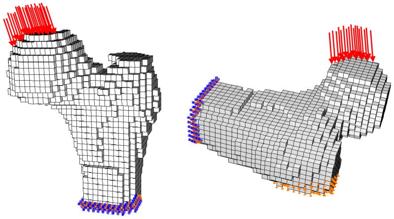
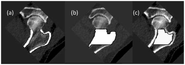
The risk of hip fracture rises rapidly with age, and is particularly high in women. This increase in fracture risk reflects both the age-related change in the risk of falling and decrements in the strength of the proximal femur. To better understand the extent to which proximal femoral density, structure and strength change with age as a function of gender, we have carried out a longitudinal analysis of proximal femoral volumetric quantitative computed tomographic (vQCT) images in men and women, analyzing changes in trabecular and cortical bone properties, and using subject-specific finite element modeling (FEM) to estimate changes in bone strength. In the AGES-Reykjavik Study vQCT scans of the hip were performed at a baseline visit in 2002-2006 and at a second visit 5.05±0.25 years later. From these, 223 subjects (111 men, 112 women, aged 68-87 years) were randomly selected. The subjects were evaluated for longitudinal changes in three bone variables assessed in a region similar to the total femur region quantified by DXA: areal bone mineral density (aBMD), trabecular volumetric bone mineral density (tBMD) and the ratio of cortical to total tissue volume (cvol/ivol). They were also evaluated for changes in bone strength using FEM models of the left proximal femur. Models were analyzed under single-limb stance loading (F(Stance)), which approximates normal physiologic loading of the hip, as well as a load approximating a fall onto the posterolateral aspect of the greater trochanter (F(Fall)). We computed five-year absolute and percentage changes in aBMD, tBMD, cvol/ivol, F(Fall) and F(Stance). The Mann-Whitney Test was employed to compare changes in bone variables between genders and the Wilcoxon Signed Rank Test was used to compare changes in bone strength between loading conditions. Multiple (linear) regression was employed to determine the association of changes in F(Fall) and F(Stance) with baseline age and five-year weight loss. Both men and women showed declines in indices of proximal femoral density and structure (aBMD: men -3.9±6.0%, women -6.1±6.2%; tBMD: men -14.8±20.3%, women -23.9±26.8%; cvol/ivol: men -2.6±4.6%, women -4.7±4.8%, gender difference: p<0.001). Both men and women lost bone strength in each loading condition (F(Stance): men -4.2±9.9%, women -8.3±8.5%; F(Fall): men -7.0±15.7%, women -12.8±13.2%; all changes from baseline p<0.0001). The gender difference in bone strength loss was statistically significant in both loading conditions (p<0.001 for F(Stance) and P<0.01 for F(Fall)) and F(Fall) was lost at a higher rate than F(Stance) in men (p<0.01) and women (p<0.0001). The gender difference in strength loss was statistically significant after adjustment for baseline age and weight loss in both loading conditions (p<0.01). In these multi-linear models, men showed increasing rates of bone loss with increasing age (F(Fall): p=0.002; F(Stance): p=0.03), and women showed increasing bone strength loss with higher degrees of weight loss (F(Stance): p=0.003). The higher loss of F(Fall) compared to F(Stance) supports previous findings in animal and human studies that the sub-volumes of bone stressed under normal physiologic loading are relatively better protected in aging. The gender difference in hip bone strength loss is consistent with the higher incidence of hip fracture among elderly women.
Copyright © 2011 Elsevier Inc. All rights reserved.
Figures
References
-
- Poole KE, Mayhew PM, Rose CM, Brown JK, Bearcroft PJ, Loveridge N, Reeve J. Changing structure of the femoral neck across the adult female lifespan. J Bone Miner Res. 2010;25:482–91. - PubMed
-
- Mayhew PM, Thomas CD, Clement JG, Loveridge N, Beck TJ, Bonfield W, Burgoyne CJ, Reeve J. Relation between age, femoral neck cortical stability, and hip fracture risk. Lancet. 2005;366:129–35. - PubMed
-
- Marshall LM, Lang TF, Lambert LC, Zmuda JM, Ensrud KE, Orwoll ES. Dimensions and Volumetric BMD of the Proximal Femur and Their Relation to Age Among Older U.S. Men. J Bone Miner Res. 2006;21:1197–206. - PubMed
-
- Riggs BL, Melton Iii LJ, 3rd, Robb RA, Camp JJ, Atkinson EJ, Peterson JM, Rouleau PA, McCollough CH, Bouxsein ML, Khosla S. Population-based study of age and sex differences in bone volumetric density, size, geometry, and structure at different skeletal sites. J Bone Miner Res. 2004;19:1945–54. - PubMed
Publication types
MeSH terms
Grants and funding
LinkOut - more resources
Full Text Sources
Medical
