

Review
doi: 10.1016/j.gde.2011.11.005.
Epub 2011 Dec 16.
Structure, assembly and reading of centromeric chromatin
Affiliations
- PMID: 22178421
- PMCID: PMC3319190
- DOI: 10.1016/j.gde.2011.11.005
Item in Clipboard
Review
Structure, assembly and reading of centromeric chromatin
Curr Opin Genet Dev.
2012 Apr.
Abstract
Centromeres are epigenetically defined chromatin domains marked by the presence of the histone H3 variant CENP-A. Here we review recent structural and biochemical work on CENP-A, and advances in understanding the mechanisms that propagate and read centromeric chromatin domains.
Copyright © 2011 Elsevier Ltd. All rights reserved.
Figures
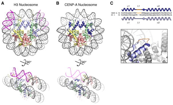
Canonical and centromeric nucleosomes. (A) Canonical H3-containing nucleosome (PBD ID 1KX5) [74], with histone H3 light blue, H4 green, H2A yellow, H2B red, and DNA gray (13 bp on each end colored magenta). (B) CENP-A-containing nucleosome (PDB ID 3AN2) [20], with CENP-A colored dark blue. Bottom: view of one DNA end, with the 13 bp of disordered DNA indicated by dashed magenta lines (figure adapted from [20]). (C) Loop 1 differences in CENP-A and H3 (adapted from [20]). Top: Sequence alignment showing the two-residue insertion in CENP-A loop 1. Bottom: closeup view of the loop 1 region, with CENP-A from [20] in dark blue and H3 from [74] in light blue. Shown in orange are CENP-A residues 79–83, which are ordered in the CENP-A monomer shown (potentially due to crystal packing interactions) and disordered in the second CENP-A monomer. An earlier CENP-A2:H42 tetramer structure also showed a distinct conformation of CENP-A loop 1 with high crystallographic B-factors indicating flexibility [21]. While mutation or truncation of loop 1 mildly affects centromere targeting of CENP-A in vivo [20], the mechanism of this defect remains unknown.
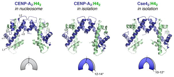
CENP-A2/Cse42:H42 tetramer structures, in the context of the full nucleosome (left) (PDB ID 3AN2) [20], and in isolation (middle: human CENP-A2:H42 (PDB ID 3NQJ) [21], right: budding yeast (K. lactis) Cse42:H42 (PDB ID 2YFW) [26]), colored as in Figure 1. Secondary structure elements of one CENP-A monomer are labeled (left panel). Both tetramer structures determined without bound H2A:H2B and DNA show compaction of the tetramer by 10–14°, relative to the conformation of both the H32:H42 and CENP-A2:H42 tetramers in the context of the full nucleosome (illustration adapted from [21]).
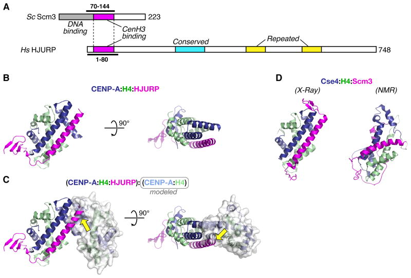
Structural analysis of the CENP-A chaperone HJURP/Scm3. (A) Diagram of S. cerevisiae Scm3 and H. sapiens HJURP. Conserved domains identified in [34] are shown: CENP-A/Cse4 binding region (Scm3 residues 90–142, HJURP residues 16–68) magenta, HJURP conserved domain (228–304) cyan, HJURP repeated regions (411–462 and 556–608) yellow. The DNA-binding region of Scm3 (1–113) identified by Xiao et al [23] is shown in gray. Constructs used for x-ray crystallography are indicated by thick lines. (B) Side and top views of the CENP-A:H4:HJURP trimer structure (PDB ID 3R45) [36], with HJURP shown in magenta. (C) The CENP-A:H4:HJURP trimer aligned with a second CENP-A:H4 dimer (molecular surface displayed), showing how HJURP partially occludes the CENP-A:CENP-A interface in the tetramer (yellow arrows). (D) Two structures of the budding-yeast Cse4:H4:Scm3 trimer. Left: structure obtained by X-ray crystallography of the proteins from K. lactis (PDB ID 2YFV) [26]. Right: structure obtained by NMR of the S. cerevisiae proteins (PDB ID 2L5A) [37]. All structures are oriented the same relative to the left-hand copy of CENP-A/Cse4 in Figure 2.
References
-
- Choo KH. Centromerization. Trends Cell Biol. 2000;10:182–188. - PubMed
-
- Maddox PS, Oegema K, Desai A, Cheeseman IM. “Holo”er than thou: chromosome segregation and kinetochore function in C. elegans. Chromosome Res. 2004;12:641–653. - PubMed
-
- Lowell JE, Cross GA. A variant histone H3 is enriched at telomeres in Trypanosoma brucei. J Cell Sci. 2004;117:5937–5947. - PubMed
Publication types
MeSH terms
Substances
Grants and funding
LinkOut - more resources
Full Text Sources
