. 2012 Feb 3;415(5):900-17.
doi: 10.1016/j.jmb.2011.12.011. Epub 2011 Dec 9.

Inhibiting the nucleation of amyloid structure in a huntingtin fragment by targeting α-helix-rich oligomeric intermediates

Affiliations

Inhibiting the nucleation of amyloid structure in a huntingtin fragment by targeting α-helix-rich oligomeric intermediates

Rakesh Mishra et al. J Mol Biol. .

Abstract

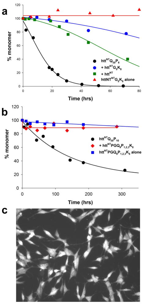
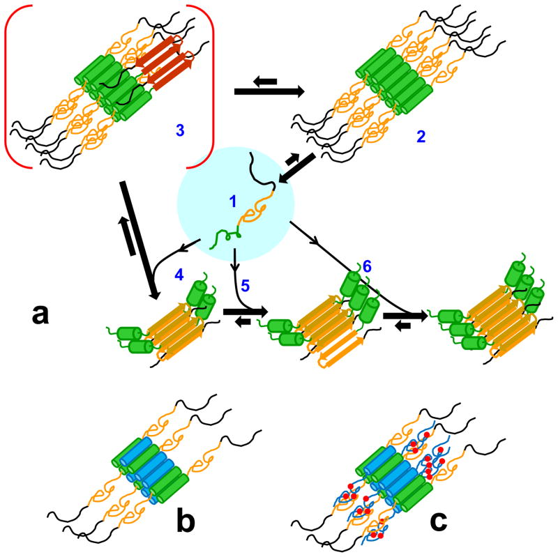
Although oligomeric intermediates are transiently formed in almost all known amyloid assembly reactions, their mechanistic roles are poorly understood. Recently, we demonstrated a critical role for the 17-amino-acid N-terminus (htt(NT) segment) of huntingtin (htt) in the oligomer-mediated amyloid assembly of htt N-terminal fragments. In this mechanism, the htt(NT) segment forms the α-helix-rich core of the oligomers, leaving much of the polyglutamine (polyQ) segment disordered and solvent-exposed. Nucleation of amyloid structure occurs within this local high concentration of disordered polyQ. Here we demonstrate the kinetic importance of htt(NT) self-assembly by describing inhibitory htt(NT)-containing peptides that appear to work by targeting nucleation within the oligomer fraction. These molecules inhibit amyloid nucleation by forming mixed oligomers with the htt(NT) domains of polyQ-containing htt N-terminal fragments. In one class of inhibitors, nucleation is passively suppressed due to the reduced local concentration of polyQ within the mixed oligomer. In the other class, nucleation is actively suppressed by a proline-rich polyQ segment covalently attached to htt(NT). Studies with D-amino acid and scrambled sequence versions of htt(NT) suggest that inhibition activity is strongly linked to the propensity of inhibitory peptides to make amphipathic α-helices. Htt(NT) derivatives with C-terminal cell-penetrating peptide segments also exhibit excellent inhibitory activity. The htt(NT)-based peptides described here, especially those with protease-resistant d-amino acids and/or with cell-penetrating sequences, may prove useful as lead therapeutics for inhibiting the nucleation of amyloid formation in Huntington's disease.


Figures

Figure 1
Figure 1
Kinetic effects of httNT on aggregation of polyQ-containing peptides by the sedimentation assay. (a) Aggregation of 23.3 μM httNTQ37P10K2 ( formula image
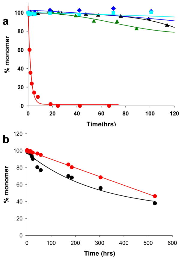
); 25.2 μM SWQ37P10K2 ( formula image); 25.6 μM K2Q35P10K2 (▴); 7.5 μM K2Q41K2 alone ( formula image); and 7.0 μM K2Q41K2 plus 17.4 μM httNT ( formula image
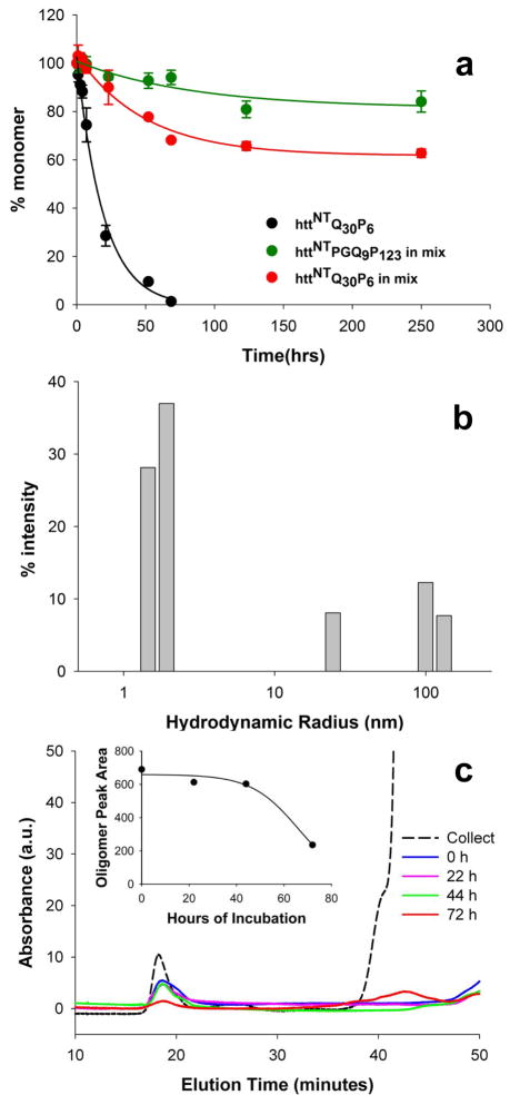
). (b) Aggregation of 18.8 μM of the peptide ESLKSF-Q35-PPPSKETAAAKFERQHMDS incubated alone (●) or with 29.6 μM httNT ( formula image).
Figure 2
Figure 2
HttNT inhibition of aggregation. (a, b) Incubation of 9 μM httNTQ30P6K2 with different molar ratios of httNT to httNTQ30P6K2: 0.05:1 (○); 0.16:1 (▴); 0.43:1 (▵); 0.89:1 (◆). (a) Soluble httNTQ30P6K2 after centrifugation; control incubation of httNTQ30P6K2 without inhibitor (●); (b) Soluble httNT inhibitor after centrifugation; control incubation of httNT alone (●); (c) Effect of higher ratios and multiple additions of httNT on httNTQ30P6K2 aggregation: 7.0 μM httNTQ30P6K2 alone (●); 5.33 μM httNTQ30P6K2 with either 7.5 μM httNT (i.e., ~ 1.4 fold molar excess) (○) or 13.3 μM httNT (i.e., ~ 2.5 fold molar excess) (▵) added at t = 0; 5.33 μM httNTQ30P6K2 plus 7.5 μM httNT reaction in which fresh, additional aliquots of httNT were added at 7 hrs (11.2 μM), 24 hrs (7.0 μM), 31 hrs (7.3 μM), and 68 hrs (6.4 μM) (■). Peptides were incubated at 37 °C in PBS, with aggregation monitored by the sedimentation assay.
Figure 3
Figure 3
The SEC profile of an incubated mixture of httNTQ30P6K2 (F17W) and httNT was analyzed by LC-MS. The peak eluting at ~ 32 mins gave the HPLC profile shown in part B, and the mass spectrum at the intensity peak (inset) gave a mass of 6697 Da consistent with httNTQ30P6K2 (F17W). The peak eluting at ~49 mins gave the HPLC profile shown in Part C, and the mass spectrum at the intensity peak (inset) gave a mass of 1972.8 Da, consistent with httNT. The absence of a SEC peak containing both peptides indicates that there is no significant low molecular weight complex formation under these conditions.
Figure 4
Figure 4
Electron micrographs of aggregates of htt N-terminal fragments. Aggregates from HttNTQ30P6K2 alone incubated in PBS at 37 °C and sampled at 0 hrs (a), 15 min (b), 5.5 hrs (c), 24 hrs (d), 48 hrs (e), and 100 hrs (f). HttNTQ30P6 incubated with httNT in 1:1 ratio in PBS, 37 °C and sampled at 3 hrs (g, h), 5.5 hrs (i, j), 24 hrs (k, l), 48 hrs (m) and 72 hrs (n). httNTPGQ9P1,2,3 incubated alone for 120 hrs (o), httNTQ30P6K2 and httNTPGQ9P1,2,3 co-incubated in 1:4 ratio in PBS, 37 °C for 120 hrs (p). Scale bar represents 50 nm.
Figure 5
Figure 5
Spectroscopic probes of aggregate structure. (a) Tryptophan fluorescence spectra of isolated and resuspended aggregates from the following reactions: monomeric httNTQ30P6K2 (F17W), λem = 355 nm (black); aggregated httNTQ30P6K2 (F17W) alone, λem = 347.5 nm, (green); httNT plus httNTQ30P6K2 (F17W), λem = 349 nm, (magenta); httNTQ3 (F17W) inhibitor plus httNTQ30P6K2, λem = 337 nm, (blue); httNTPGQ9P1,2,3 (F17W) plus httNTQ30P6K2, λem = 347.5 nm, (orange). (b) Time course of the Trp fluorescence peak shift in isolated aggregates when the httNTQ3 (F17W) inhibitor is incubated with httNTQ30P6K2 at a 1.1:1 ratio; (c) Second deriviative FTIR spectra of aggregates isolated at about 100 hrs from incubation reactions of httNT (black), httNTQ30P6K2 (red), and a reaction with a 1:1 mixture of httNT and httNTQ30P6K2 (blue); (d) primary FTIR spectra of aggregates generated upon incubation of either all-L httNT (blue), all-D httNT (red), and retro-inverso httNT (green).
Figure 6
Figure 6
Abilities of approximately equimolar httNT analogs to inhibit aggregation of htt N-terminal fragments in PBS at 37 °C, as determined by a sedimentation assay. (a) inhibition of 7.6 μM httNTQ30P10K2 aggregation by all-L httNT (15 μM), all-D httNT (11.1 μM) and retroinverso-httNT (9.5 μM) (b, c) inhibition of 5–10 μM httNTQ30P6K2 aggregation by 1:1 molar amounts of wild type and scrambled httNTQ sequences.
Figure 7
Figure 7
Scatterplot analyses of inhibitory activities of wild type httNT and scrambled peptides (Table 2). (a) aggregation inhibition vs. AGADIR α-helix propensity (linear fit; R2 = 0.80); (b) aggregation inhibition vs. hydrophobic moment (μH) score (exponential fit; R2 = 0.88); (c) aggregation inhibition vs. normalized μH values determined by multiplying the hydrophobic moment times the measured % α-helix as determined by CD spectroscopy (three parameter exponential fit; R2 = 0.70) (see Table 2). In each plot the position of the httNT wild type sequence is in red.
Figure 8
Figure 8
Properties of httNTPGQ9P1,2,3. (a) inhibition httNTQ30P6K2 aggregation by equimolar httNTPGQ9P1,2,3; (b) DLS of httNTPGQ9P1,2,3 incubated alone for three hrs; (c) SEC analysis of oligomer assembly and disassembly of httNTPGQ9P1,2,3. 300 μM peptide was incubated for 24 hrs, then separated by SEC in PBS running buffer (dashed line). The oligomer peak (~18 mins) was collected and incubated at 37°C, then analyzed at the times shown. Between 44 and 72 hrs a substantial portion of oligomer (~18 mins) dissociates to monomer (~42 mins).
Figure 9
Figure 9
Properties of cell penetrating peptide versions of inhibitors. (a) Inhibition of httNTQ30P6K2 aggregation by httNTG5K8 and controls; (b) Inhibition of httNTQ30P10K2 aggregation by httNT PGQ9P1,2,3 K8 and controls; c) Cytosolic delivery of httNTG4CK8-Alexa633 into SHSY5Y cells (Methods). After 10 mins exposure to peptide plus 1-pyrenebutyric acid, cells were washed extensively with PBS and imaged.
Figure 10
Figure 10
α-Helix rich molecules in htt N-terminal fragment aggregation and its inhibition. (a) A model for nucleated growth in htt N-terminal fragment aggregation (modified from Jayaraman et al., Ms. submitted). When the htt N-terminal fragment monomer (a1: httNT, green; polyQ, orange; Pro-rich domain, black) assembles into the oligomeric intermediate (a2), the httNT segment takes on α-helix structure whose packing with other httNT segments into a helical bundle stabilizes the oligomer. As a consequence, the disordered polyQ chains are brought together closely in space within the oligomer, which lowers the barrier to formation of nuclei with amyloid structural elements (a3, polyQ extended chain, red). Monomers present at this stage facilitate elongation into growing amyloid fibrils (a4, a5, a6). In the final stages of amyloid growth, as the monomer pool is depleted, oligomers that have not undergone nucleation dissociate (a2 -> a1) to generate more monomers to support fibril elongation. (b) structural model for the inhibition complex (oligomer) between httNT (blue cylinders) and an htt N-terminal fragment; (c) structural model for the inhibition complex between an htt N-terminal fragment and a C-terminally extended httNT inhibitor, such as httNTPGQ9P1,2,3 or httNTPGQ9P1,2,3K8 (blue cylinders = httNT helices; blue lines = PGQ9 sequence; red circles = additional Pro residues (see Table 1)).

Similar articles

Cited by

References

    1. Zuccato C, Valenza M, Cattaneo E. Molecular mechanisms and potential therapeutical targets in Huntington’s disease. Physiol Rev. 2010;90:905–81. - PubMed
    1. Wilburn B, Rudnicki DD, Zhao J, Weitz TM, Cheng Y, Gu XF, Greiner E, Park CS, Wang N, Sopher BL, La Spada AR, Osmand A, Margolis RL, Sun YE, Yang XW. An antisense CAG repeat transcript at JPH3 locus mediates expanded polyglutamine protein toxicity in Huntington’s disease-like 2 mice. Neuron. 2011;70:427–440. - PMC - PubMed
    1. Bates GP, Benn C. The polyglutamine diseases. In: Bates GP, Harper PS, Jones L, editors. Huntington’s Disease. Oxford University Press; Oxford, U.K: 2002. pp. 429–472.
    1. Sathasivam K, Lane A, Legleiter J, Warley A, Woodman B, Finkbeiner S, Paganetti P, Muchowski PJ, Wilson S, Bates GP. Identical oligomeric and fibrillar structures captured from the brains of R6/2 and knock-in mouse models of Huntington’s disease. Human Molecular Genetics. 2010;19:65–78. - PMC - PubMed
    1. Scherzinger E, Sittler A, Schweiger K, Heiser V, Lurz R, Hasenbank R, Bates GP, Lehrach H, Wanker EE. Self-assembly of polyglutamine-containing huntingtin fragments into amyloid-like fibrils: implications for Huntington’s disease pathology. Proc Natl Acad Sci U S A. 1999;96:4604–9. - PMC - PubMed

Publication types

MeSH terms