. 2012 Apr;142(4):855-864.e8.
doi: 10.1053/j.gastro.2011.12.004. Epub 2011 Dec 13.

Field cancerization in the intestinal epithelium of patients with Crohn's ileocolitis

Affiliations

Field cancerization in the intestinal epithelium of patients with Crohn's ileocolitis

Susan Galandiuk et al. Gastroenterology. 2012 Apr.

Abstract

Background & aims: Tumors that develop in patients with Crohn's disease tend be multifocal, so field cancerization (the replacement of normal cells with nondysplastic but tumorigenic clones) might contribute to intestinal carcinogenesis. We investigated patterns of tumor development from pretumor intestinal cell clones.

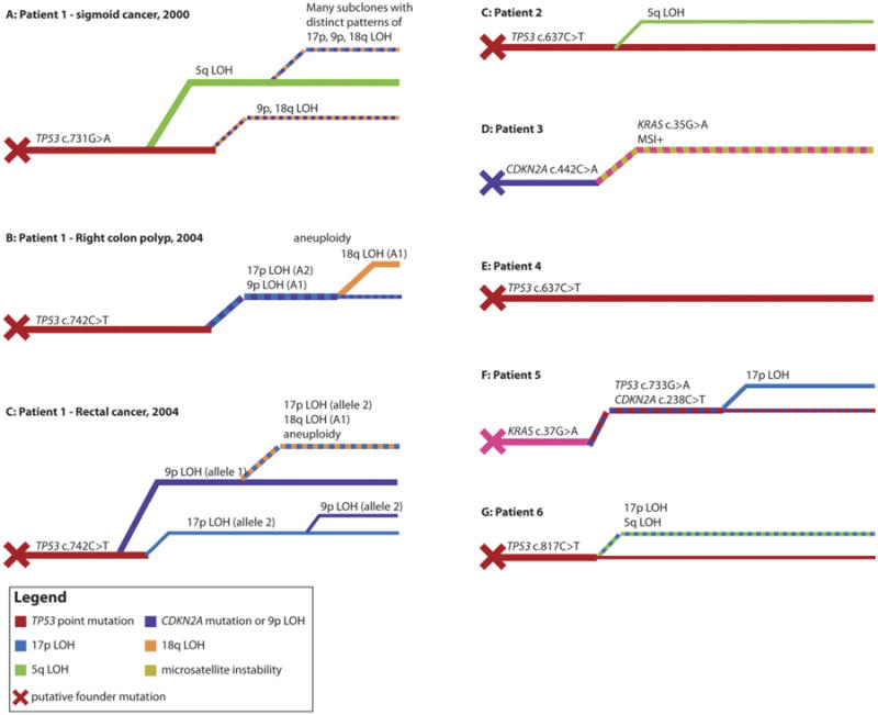
Methods: We performed genetic analyses of multiple areas of intestine from 10 patients with Crohn's disease and intestinal neoplasia. Two patients had multifocal neoplasia; longitudinal sections were collected from 3 patients. Individual crypts were microdissected and genotyped; clonal dependency analysis was used to determine the order and timing of mutations that led to tumor development.

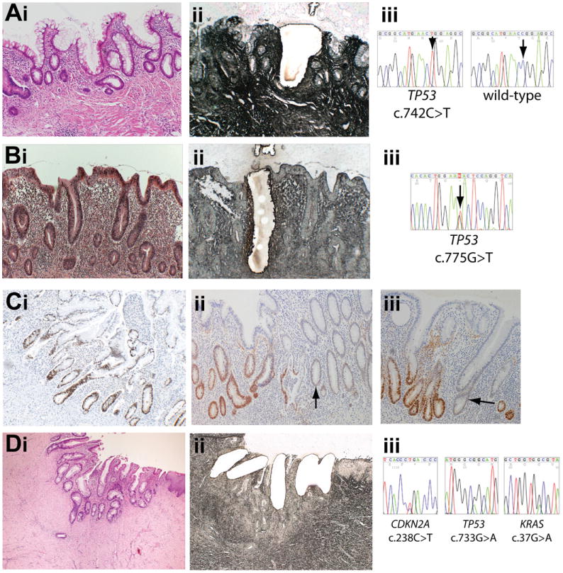
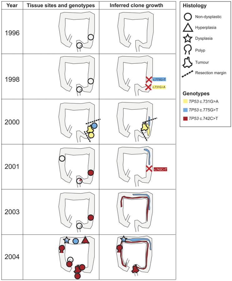
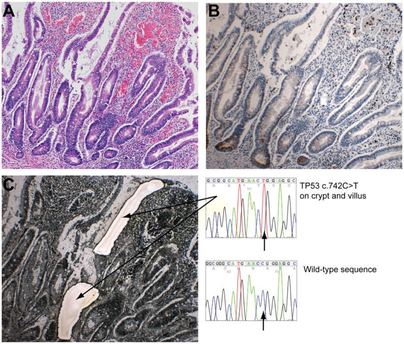
Results: The same mutations in KRAS, CDKN2A(p16), and TP53 that were observed in neoplasias were also present in nontumor, nondysplastic, and dysplastic epithelium. In 2 patients, carcinogenic mutations were detected in nontumor epithelium 4 years before tumors developed. The same mutation (TP53 p.R248W) was detected at multiple sites along the entire length of the colon from 1 patient; it was the apparent founder mutation for synchronous tumors and multiple dysplastic areas. Disruption of TP53, CDKN2A, and KRAS were all seen as possible initial events in tumorigenesis; the sequence of mutations (the tumor development pathway) differed among lesions.

Conclusions: Pretumor clones can grow extensively in the intestinal epithelium of patients with Crohn's disease. Segmental resections for neoplasia in patients with Crohn's disease might therefore leave residual pretumor disease, and dysplasia might be an unreliable biomarker for cancer risk. Characterization of the behavior of pretumor clones might be used to predict the development of intestinal neoplasia.


Conflict of interest statement

Conflicts of interest: The authors disclose no conflicts.

Figures

Figure 1
Figure 1
Mutations present in nontumor tissue. (A) (i) H&E stain (original magnification 100×) of nondysplastic (hyperplastic) mucosa showing crypt distortion with increased inflammatory cells and intraepithelial neutrophils in the transverse colon in patient 1 in 2004 and (ii) serial methylene green–stained PALM laser capture slide showing microdissected crypt. (iii) Sequencing shows the crypt contains a TP53 c.742C>T mutation. (B) (i) H&E stain (original magnification 100×) of nondysplastic (hyperplastic) mucosa with a marked increase in the inflammatory cells in the lamina propria and cryptitis in the resection margin of the sigmoid cancer resected from patient 1 in 2000 with (ii) serial PALM slide showing microdissection. (iii) Sequencing shows the crypt contains a TP53 c.775G>T mutation. (C) p53 immunohistochemistry on a nondysplastic colon resection specimen from patient 1 (original magnification 100×). (i) Patches of crypts with nuclear accumulation of p53 protein were observed, frequently demarked by odd p53-negative or low-expressing crypts (ii and iii) (arrows). (D) (i) H&E stain (original magnification 40×) of inflammatory atypia within cells in a perianal fistula from patient 5 four years before tumor growth. (ii) Laser capture slide (original magnification 100×). (iii) TP53, CDKN2A, and KRAS mutations in each crypt.
Figure 2
Figure 2
Longitudinal analysis showing clone spread in patient 1. Tissue collected before 2000 contained no widespread detected genetic abnormalities. A c.731G>A TP53 mutation was first detected throughout a sigmoid cancer in 2000 and within the resection margins, but not in later samples, suggesting the mutant arose in the sigmoid after the biopsy in 1998 and was completely removed by the cancer resection. A second c.775G>T TP53 was also found in the resection margin of the sigmoid cancer. This second clone had spread to the transverse colon by 2004. A third TP53 c.742C>T mutation was first detected in sigmoid and rectal biopsy specimens from 2001. By 2004, the mutant TP53 c.742C>T clone was present in every major segment of the colon and also in the terminal ileum and in multifocal neoplasia. Shapes indicate the predominant pathological diagnosis at the site, and color indicates the predominant TP53 genotype (see key).
Figure 3
Figure 3
Presence of the same TP53 mutation in functional small intestinal and colon crypts. (A) H&E section showing inflamed terminal ileum mucosa with crypt/villous distortion and an increase in inflammatory cells and intraepithelial neutrophils. (B) Serial section stained for lysozyme indicating the presence of functional Paneth cells at the small intestinal crypt base. (C) Serial laser capture slide stained with methylene green showing separately microdissected crypt and villus. Genotyping revealed the same TP53 c.742C>T was present in both the crypt and villus. This same mutation was found in colon tissue collected at the same and previous times. Original magnification 100× for all micrographs.
Figure 4
Figure 4
Phylogenetic trees of neoplasia development. (A) Phylogenetic tree of sigmoid cancer resected in patient 1 in 2000. Tumor growth was preceded by the acquisition of a TP53 c.731G>A mutation, because this mutation was found in the cancer and its resection margin. A clone with 5q LOH was dominant in the cancer, and a single crypt showed 9p and 18q LOH but no 5q LOH. Numerous small subclones with distinct patterns of 9p, 18q, and 17p LOH were found within the 5q subclone. This frequent LOH could be attributable to the cancer containing an aneuploid clone, which was not tested for in this cancer. (B) Phylogenetic tree of the ascending colon adenoma resected in patient 1 in 2004. The cancer developed from the preneoplastic clone distinguished by the TP53 c.742C>T mutation, and subsequent carcinogenesis involved the sequential acquisition of 17p, 9p, and then 18q LOH. Unlike the previous cancer in patient 1, no 5q LOH was detected in the lesion in 2004. (C–G) Phylogenetic trees for tumors from patients 1–6. Most lesions were initiated by a TP53 mutant clone, with the exceptions of patient 3, where the putative founder clone was a CDKN2A (p16) mutant, and patient 5, where the founder clone was a KRAS mutant. The inferred sequence of subsequent mutations and LOH events was different in every lesion. Colors indicate genotypes (see key), and line thicknesses are indicative of relative clone abundance.

References

    1. Laukoetter MG, Mennigen R, Hannig CM, et al. Intestinal cancer risk in Crohn's disease: a meta-analysis. J Gastrointest Surg. 2011;15:576–583. - PubMed
    1. Hemminki K, Li X, Sundquist J, et al. Cancer risks in Crohn disease patients. Ann Oncol. 2009;20:574–580. - PubMed
    1. von Roon AC, Reese G, Teare J, et al. The risk of cancer in patients with Crohn's disease. Dis Colon Rectum. 2007;50:839–855. - PubMed
    1. Canavan C, Abrams KR, Mayberry J. Meta-analysis: colorectal and small bowel cancer risk in patients with Crohn's disease. Aliment Pharmacol Ther. 2006;23:1097–1104. - PubMed
    1. Gyde SN, Prior P, Macartney JC, et al. Malignancy in Crohn's disease. Gut. 1980;21:1024–1029. - PMC - PubMed

Publication types

MeSH terms