

Damage to mitochondrial complex I during cardiac ischemia reperfusion injury is reduced indirectly by anti-anginal drug ranolazine
- PMID: 22178605
- PMCID: PMC3269517
- DOI: 10.1016/j.bbabio.2011.11.021
Damage to mitochondrial complex I during cardiac ischemia reperfusion injury is reduced indirectly by anti-anginal drug ranolazine
Abstract
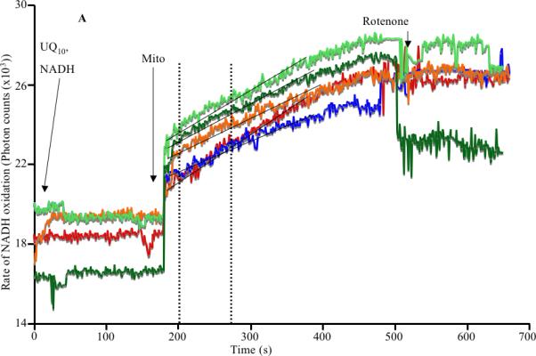
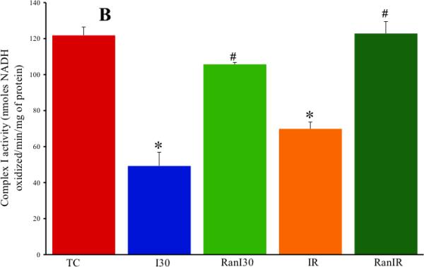
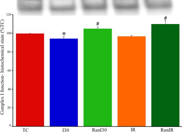
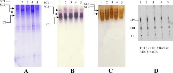
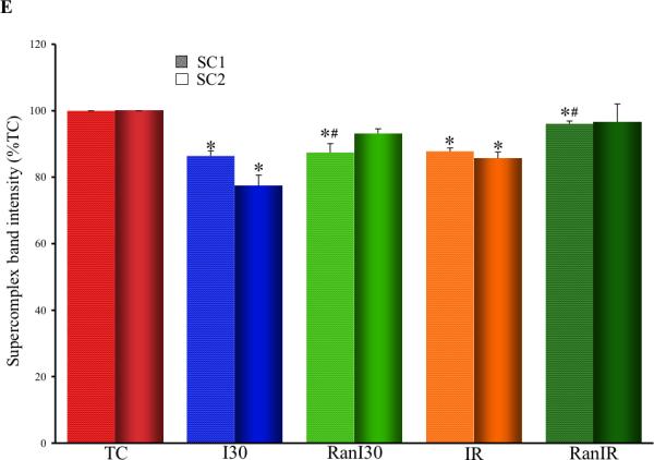
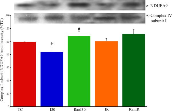
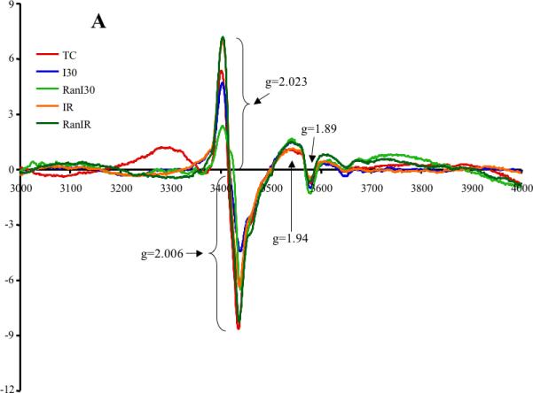
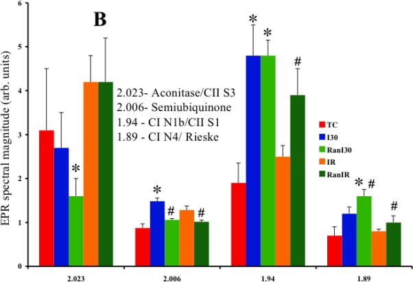
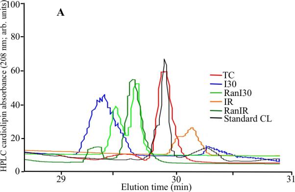
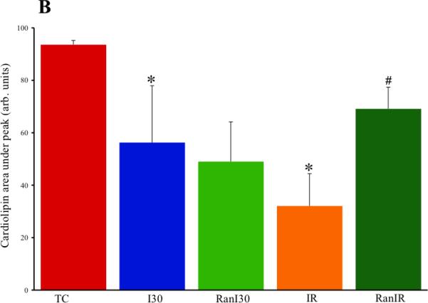
Ranolazine, an anti-anginal drug, is a late Na(+) channel current blocker that is also believed to attenuate fatty acid oxidation and mitochondrial respiratory complex I activity, especially during ischemia. In this study, we investigated if ranolazine's protective effect against cardiac ischemia/reperfusion (IR) injury is mediated at the mitochondrial level and specifically if respiratory complex I (NADH Ubiquinone oxidoreductase) function is protected. We treated isolated and perfused guinea pig hearts with ranolazine just before 30 min ischemia and then isolated cardiac mitochondria at the end of 30 min ischemia and/or 30 min ischemia followed by 10 min reperfusion. We utilized spectrophotometric and histochemical techniques to assay complex I activity, Western blot analysis for complex I subunit NDUFA9, electron paramagnetic resonance for activity of complex I Fe-S clusters, enzyme linked immuno sorbent assay (ELISA) for determination of protein acetylation, native gel histochemical staining for respiratory supercomplex assemblies, and high pressure liquid chromatography for cardiolipin integrity; cardiac function was measured during IR. Ranolazine treated hearts showed higher complex I activity and greater detectable complex I protein levels compared to untreated IR hearts. Ranolazine treatment also led to more normalized electron transfer via Fe-S centers, supercomplex assembly and cardiolipin integrity. These improvements in complex I structure and function with ranolazine were associated with improved cardiac function after IR. However, these protective effects of ranolazine are not mediated by a direct action on mitochondria, but rather indirectly via cytosolic mechanisms that lead to less oxidation and better structural integrity of complex I.
Copyright © 2011 Elsevier B.V. All rights reserved.
Figures
Similar articles
-
Ranolazine reduces Ca2+ overload and oxidative stress and improves mitochondrial integrity to protect against ischemia reperfusion injury in isolated hearts.Pharmacol Res. 2011 Oct;64(4):381-92. doi: 10.1016/j.phrs.2011.06.018. Epub 2011 Jun 29. Pharmacol Res. 2011. PMID: 21741479 Free PMC article.
-
Protective effects of ranolazine and propranolol, alone or combined, on the structural and functional alterations of cardiomyocyte mitochondria in a pig model of ischemia/reperfusion.Fundam Clin Pharmacol. 2014 Jun;28(3):257-67. doi: 10.1111/fcp.12033. Epub 2013 Apr 22. Fundam Clin Pharmacol. 2014. PMID: 23607936
-
The antianginal agent ranolazine is a weak inhibitor of the respiratory complex I, but with greater potency in broken or uncoupled than in coupled mitochondria.Biochem Pharmacol. 1995 Nov 9;50(10):1599-606. doi: 10.1016/0006-2952(95)02042-x. Biochem Pharmacol. 1995. PMID: 7503762
-
Ranolazine, a partial fatty acid oxidation inhibitor, its potential benefit in angina and other cardiovascular disorders.Recent Pat Cardiovasc Drug Discov. 2007 Jan;2(1):35-9. doi: 10.2174/157489007779606095. Recent Pat Cardiovasc Drug Discov. 2007. PMID: 18221101 Review.
-
Anti-anginal and anti-ischemic effects of late sodium current inhibition.Cardiovasc Drugs Ther. 2013 Feb;27(1):69-77. doi: 10.1007/s10557-012-6431-z. Cardiovasc Drugs Ther. 2013. PMID: 23247666 Free PMC article. Review.
Cited by
-
Safety and Efficacy of Ranolazine for the Treatment of Chronic Angina Pectoris.Clin Med Insights Ther. 2013 Jan 15;2013(5):1-14. doi: 10.4137/CMT.S7824. Clin Med Insights Ther. 2013. PMID: 24574825 Free PMC article.
-
Conformational change of mitochondrial complex I increases ROS sensitivity during ischemia.Antioxid Redox Signal. 2013 Nov 1;19(13):1459-68. doi: 10.1089/ars.2012.4698. Epub 2013 Mar 29. Antioxid Redox Signal. 2013. PMID: 23419200 Free PMC article.
-
The Role of Mitochondrial Reactive Oxygen Species in Cardiovascular Injury and Protective Strategies.Oxid Med Cell Longev. 2016;2016:8254942. doi: 10.1155/2016/8254942. Epub 2016 Apr 21. Oxid Med Cell Longev. 2016. PMID: 27200148 Free PMC article. Review.
-
Cardiac mitochondria and reactive oxygen species generation.Circ Res. 2014 Jan 31;114(3):524-37. doi: 10.1161/CIRCRESAHA.114.300559. Circ Res. 2014. PMID: 24481843 Free PMC article. Review.
-
Early Effects of Prolonged Cardiac Arrest and Ischemic Postconditioning during Cardiopulmonary Resuscitation on Cardiac and Brain Mitochondrial Function in Pigs.Resuscitation. 2017 Jul;116:8-15. doi: 10.1016/j.resuscitation.2017.03.033. Epub 2017 Apr 10. Resuscitation. 2017. PMID: 28408349 Free PMC article.
References
-
- Aldakkak M, Stowe DF, Chen Q, Lesnefsky EJ, Camara AK. Inhibited mitochondrial respiration by amobarbital during cardiac ischaemia improves redox state and reduces matrix Ca2+ overload and ROS release. Cardiovasc. Res. 2008;77:406–415. - PubMed
-
- Paradies G, Petrosillo G, Pistolese M, Di Venosa N, Federici A, Ruggiero FM. Decrease in mitochondrial complex I activity in ischemic/reperfused rat heart: involvement of reactive oxygen species and cardiolipin. Circ. Res. 2004;94:53–59. - PubMed
-
- Fry M, Green DE. Cardiolipin requirement for electron transfer in complex I and III of the mitochondrial respiratory chain. J. Biol. Chem. 1981;256:1874–1880. - PubMed
Publication types
MeSH terms
Substances
Grants and funding
LinkOut - more resources
Full Text Sources
Other Literature Sources
Miscellaneous
