Review
. 2012 Mar 15;519(2):210-22.
doi: 10.1016/j.abb.2011.11.021. Epub 2011 Dec 8.

Metal site occupancy and allosteric switching in bacterial metal sensor proteins

Affiliations
Review

Metal site occupancy and allosteric switching in bacterial metal sensor proteins

Alfredo J Guerra et al. Arch Biochem Biophys. .

Abstract

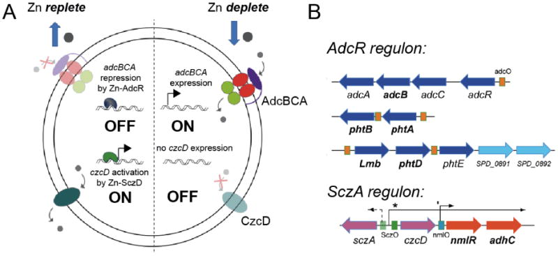
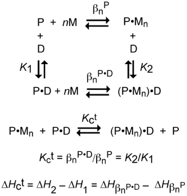
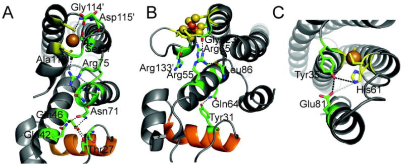
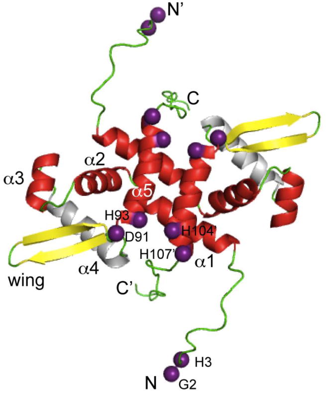
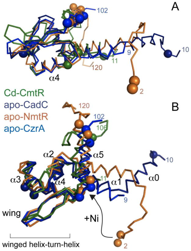
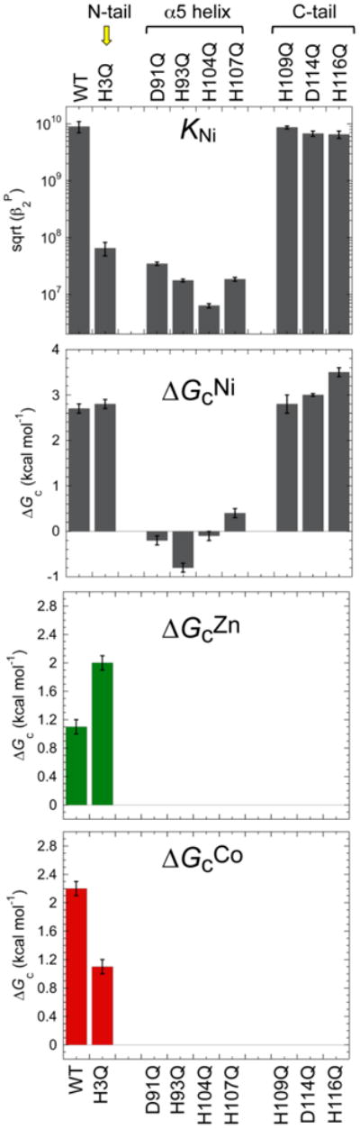
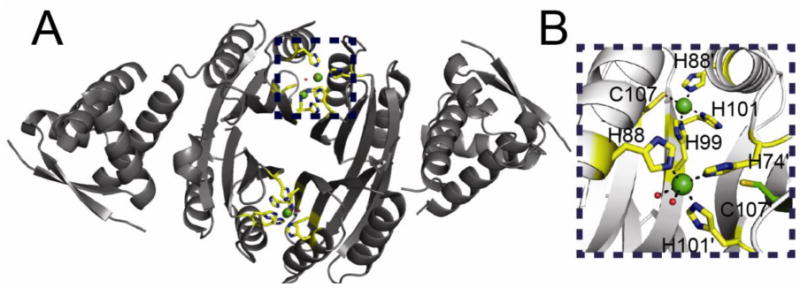
All prokaryotes encode a panel of metal sensor or metalloregulatory proteins that govern the expression of genes that allows an organism to quickly adapt to toxicity or deprivation of both biologically essential transition metal ions, e.g., Zn, Cu, Fe, and heavy metal pollutants. As such, metal sensor proteins can be considered arbiters of intracellular transition metal bioavailability and thus potentially control the metallation state of the metalloproteins in the cell. Metal sensor proteins are specialized allosteric proteins that regulate transcription as a result direct binding of one or two cognate metal ions, to the exclusion of all others. In most cases, the binding of the cognate metal ion induces a structural change in a protein oligomer that either activates or inhibits operator DNA binding. A quantitative measure of the degree to which a particular metal drives metalloregulation of operator DNA-binding is the allosteric coupling free energy, ΔGc. In this review, we summarize recent work directed toward understanding metal occupancy and metal selectivity of these allosteric switches in selected families of metal sensor proteins and examine the structural origins of ΔGc in the functional context a thermodynamic "set-point" model of intracellular metal homeostasis.


Figures

Fig. 1
Fig. 1
Regulation of zinc homeostasis in Streptococcus pneumonaie. AdcR (adhesin competence repressor) and SczA (streptococcal czcD activator) are uptake and efflux regulators of zinc and thus control zinc availability in the cytoplasm of Sp. AdcR is a member of the MarR family of bacterial repressors [11] and SczA is a TetR (tetracycline repressor) ortholog [9].
Fig. 2
Fig. 2
The set-point model of metal homeostasis [1,22,23]. Representation of how the metal affinity (KZn) of a pair of Zn(II)-dependent regulators, AdcR [11] and SczA (G. Campanello and D. Giedroc, unpublished), can effectively buffer the concentration of Zn(II) in the cell, where 1/KZn defines a fractional repression/derepression of 0.5. This set-point model is based on the assumption that metal sensor proteins quickly equilibrate and effectively scan the cytoplasm for bioavailable metal. Dashed curve, Superposition of negative homotropic cooperativity of metal binding to a dimeric regulator on the set-point model, with KZn1=1012 M−1, KZn2 =5×10¹⁰ M−1, or a degree of negative cooperativity observed experimentally in BsZur [52] and SaCzrA [48]. As can be seen, negative cooperativity of zinc binding to an uptake regulator has the effect of increasing the range of [Zn]f over which metal-responsive repression is observed [52]. The same would be true for an efflux regulator, e.g., SaCzrA, on the derepression arm (SczA) of this set-point model. The competitiveness of a particular metal follows the Irving-Williams series for divalent cations [24,25,133]; monovalent Cu(I) is known to bind to chelates far more tightly than Zn(II) [1,18].
Fig. 3
Fig. 3
General coupled thermodynamic cycle that describes the relationship between the four allosteric states of a homodimeric or homotetrameric protein P in equilibrium with a total number of n metal ions (M) per oligomer and its DNA operator (D). βn is the overall equilibrium constant for the binding of n metal ions to the oligomer. The coupling constant is given by Kct, and is defined by the disproportion equilibrium shown.
Fig. 4
Fig. 4
Surface representation of four Zn(II)-dependent transcriptional regulators structurally characterized. These are E. coli ZntR (A) [18], S. coelicolor Zur(B) [66], S. aureus CzrA (C) [68], and S. pneumoniae AdcR (D) [67]. Metal ions are represented as spheres and metal binding ligands are represented as sticks with carbon atoms colored in yellow. Metal binding sites in each protein have high solvent accessibility.
Fig. 5
Fig. 5
Close-up view of the regulatory metal binding sites of E. coli ZntR (A), S. coelicolor Zur(B), S. aureus CzrA (C), and S. pneumoniae AdcR (D). Note that three of the four Zn(II) regulators harbor two metal binding sites that are within 10 Å of one another, either as part of a binuclear metal center consisting of two shared ligands, as in E. coli ZntR (panel A) [18] or in an amino acid sequence as is the case in S. coelicolor Zur [65,66] and S. pneumoniae AdcR [67] in panels B and D, respectively.
Fig. 6
Fig. 6
Illustration of multiple binding sites in the Fur family of repressor proteins. HpFur [64], ScZur [66], ScNur [75] and BsPerR [77] illustrate both the total number of different sites as well as the different coordination complexes that have evolved in a single protein family. Metal site designations are internally consistent and labeled sites 1, 2 and 3 according to the convention established for HpFur and MtZur [65] to facilitate comparisons. Sites 1, 2 and 3 in ScZur correspond to the sites previously identified at C, M and D, respectively.
Fig. 7
Fig. 7
Putative hydrogen bonding network in the allosterically activated states of EcCueR [18] (A), EcSoxR (B) [90], and MtCsoR (C) [91]. Metal binding residues are shown in stick representation with carbon atoms shaded yellow. Key residues in the allosteric hydrogen bonding pathway are also shown as sticks with carbon atoms shaded green. The DNA recognition helix of the HTH DNA binding domain of CueR and SoxR is shaded orange. A native chemical ligation experiment carried out with MtCsoR is consistent with the coupling model shown [41]; the others have not yet been tested.
Fig. 8
Fig. 8
Ribbon representation of the structure of the Ni(II) regulator MtNmtR solved by NMR spectroscopy [101] (PDB code 2LKP). Metal binding ligands are represented as purple spheres on the Cα atom of each residue. Secondary structural elements are shaded red (α-helices), yellow (β-strands), and green (loops and unstructured regions). The N-and C- terminal extensions including residues 2-16 and 109-120 are highly mobile on the ps-ns timescale and defined the limits of the unstructured terminal regions in NmtR [59]; in contrast, residues 17-108 are highly ordered.
Fig. 9
Fig. 9
Cα-cartoon superimposition of one of the two protomers of dimeric ArsR family metal sensors MtCmtR (shaded green) [102], SaCadC (dark blue) [103], MtNmtR (gold) [101] and SaCzrA (light blue) [68]. All proteins were aligned their HTH-DNA binding motifs with an r.m.s.d. of ∼1 Å for the corresponding 29 Cα atoms. Metal binding residues in each protein are highlighted as spheres on their Cα atoms. A possible movement of the N-terminal extension of MtNmtR is also shown [59].
Fig. 10
Fig. 10
Coupling free energy analysis of MtNmtR [59]. Mutations to Ni(II) binding residues in the α5 helix greatly reduce Ni(II) binding affinity, and abrogate the coupling free energy that connects Ni(II)-binding to negative regulation of DNA-binding. Mutation of H3 in the N-terminal tail, in contrast, lowers the Ni(II) affinity by the nearly the same degree as the α5 mutants, but is characterized by a near wild-type coupling free energy. However, the H3A substitution effectively switches the rank order of allosteric effectors, with this mutant recovering significant allosteric response to the non-cognate Zn(II).
Fig. 11
Fig. 11
(A) Ribbon representation of the two pairs of Ni(II) binding sites of HpNikR solved at pH 5.6 [107]. Ni(II) ions are represented as green spheres. Metal binding residues are shown in stick representation with carbon atoms shaded yellow. (B) Close up of the two binding sites along the “top” of the NikR tetramer. Metal binding residues are shown as sticks with carbon atoms colored yellow. This view illustrates the two different coordination geometries that are found in this structure. The non-liganding Cys107 which forms part of the canonical square planar site (top site) is also shown in stick representation with carbon atoms colored green.

References

    1. Reyes-Caballero H, Campanello GC, Giedroc DP. Biophys Chem. 2011;156:103–114. - PMC - PubMed
    1. Giedroc DP, Arunkumar AI. Dalton Trans. 2007;29:3107–3120. - PubMed
    1. Ma Z, Jacobsen FE, Giedroc DP. Chem Rev. 2009;109:4644–4681. - PMC - PubMed
    1. Busenlehner LS, Pennella MA, Giedroc DP. FEMS Microbiol Rev. 2003;27:131–143. - PubMed
    1. Osman D, Cavet JS. Nat Prod Rep. 2010;27:668–680. - PubMed

Publication types