. 2011 Dec 27;108(52):20992-7.
doi: 10.1073/pnas.1116034108. Epub 2011 Dec 16.

Capture and imaging of a prehairpin fusion intermediate of the paramyxovirus PIV5

Affiliations

Capture and imaging of a prehairpin fusion intermediate of the paramyxovirus PIV5

Yong Ho Kim et al. Proc Natl Acad Sci U S A. .

Abstract

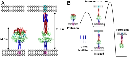
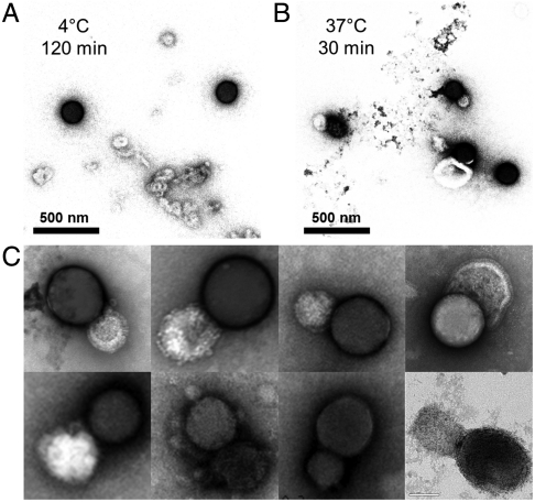
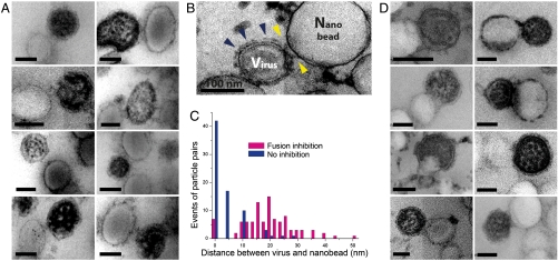
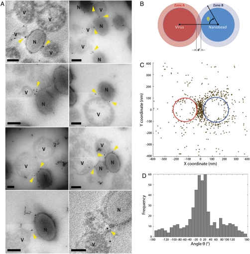
During cell entry, enveloped viruses fuse their viral membrane with a cellular membrane in a process driven by energetically favorable, large-scale conformational rearrangements of their fusion proteins. Structures of the pre- and postfusion states of the fusion proteins including paramyxovirus PIV5 F and influenza virus hemagglutinin suggest that this occurs via two intermediates. Following formation of an initial complex, the proteins structurally elongate, driving a hydrophobic N-terminal "fusion peptide" away from the protein surface into the target membrane. Paradoxically, this first conformation change moves the viral and cellular bilayers further apart. Next, the fusion proteins form a hairpin that drives the two membranes into close opposition. While the pre- and postfusion hairpin forms have been characterized crystallographically, the transiently extended prehairpin intermediate has not been visualized. To provide evidence for this extended intermediate we measured the interbilayer spacing of a paramyxovirus trapped in the process of fusing with solid-supported bilayers. A gold-labeled peptide that binds the prehairpin intermediate was used to stabilize and specifically image F-proteins in the prehairpin intermediate. The interbilayer spacing is precisely that predicted from a computational model of the prehairpin, providing strong evidence for its structure and functional role. Moreover, the F-proteins in the prehairpin conformation preferentially localize to a patch between the target and viral membranes, consistent with the fact that the formation of the prehairpin is triggered by local contacts between F- and neighboring viral receptor-binding proteins (HN) only when HN binds lipids in its target membrane.


Conflict of interest statement

The authors declare no conflict of interest.

Figures

Fig. 1.
Fig. 1.
Structures of the fusion protein in fusion states and their thermodynamics. (A) Models of the prefusion (7, 27) (left) and the prehairpin intermediate bound by the fusion inhibitor (right) show the distance expected to be observed in EM. The fusion peptide is shown in cyan, heptad repeat A in red, the globular head domain in green, heptad repeat B in dark blue (cartoon), and the TM domain in magenta. The fusion-inhibitory peptide, C1 (12), is shown in dark blue (filled cylinders). (B) Schematic diagram illustrating the free energy changes in the system. The canonical pathway moves from prefusion, to the intermediate state, through a high barrier to the postfusion state (8). Introduction of the fusion-inhibitory peptide, C1, traps the protein in the intermediate state conformation (12).
Fig. 2.
Fig. 2.
Temperature triggers viral fusion with nanobead-supported bilayers. EM images of the samples. (A) At 4 °C, nanobead-supported bilayers made up of silica nanobeads were combined with purified PIV5 particles that express the fusion (F) protein spike on their surface. At this temperature there is little association between the virus and the nanobead-supported bilayers. Dark spheres represent silica nanobead and light vesicles are viruses. Silica-supported bilayers appear darker under these conditions. (B) After warming to 37 °C for 30 min viral particles fuse to nanobead-supported bilayers. Samples in (A and B) were stained with 4% uranyl acetate. (C) EM gallery of fused virus/nanobead-supported bilayers pairs in higher magnification. PIV5 virions with a concentration of 1.0 × 1010 plaque forming units (PFU)/mL were mixed with silica nanobeads with an approximate concentration of 5 × 109 particles/mL into 100 μL total volumes and incubated at the desired temperature. The concentration of silica nanobeads was calculated from weight, assuming a 120 nm mean diameter of silica and its density of 2.2 g/cm3.
Fig. 3.
Fig. 3.
Observation of the prehairpin intermediate by thin-sectioning and EM. EM gallery of viral particles and PS nanobead-supported bilayers made up of polystyrene beads from thin sectioned samples (A) at 42 °C in the presence of the fusion-inhibitory peptide C-1 and (B) in the absence of the fusion-inhibitory peptide, C-1, under the same condition as in (A). Under these staining conditions viral particles are darker than the PS nanobeads. (B) Blue arrows show the putative prefusion state in the absence of the fusion-inhibitory peptide C-1, while yellow arrows show putative prehairpin intermediates. (C) Distance distribution between the edge of the viral particle and nanobead with and without fusion inhibitor at 42 °C. Note the peak near 20 nm for the inhibited state. (D) EM gallery of viral particles fusing with nanobead-supported bilayers at 42 °C in the absence of fusion-inhibitory peptides C-1. PIV5 virions with a concentration of 1.0 × 1010 plaque forming units/mL were mixed with amino polystyrene nanobeads with a concentration of 5 × 1010 particles/mL. The faint appearance of sections in EM images is an artifact arising from four independent quadrants in the camera used for image capture.
Fig. 4.
Fig. 4.
Immuno-gold labeling of the fusion inhibitor shows it is concentrated between the viral particles and nanobead-supported bilayers in the trapped state. (A) Gallery of thin sectioned EM images of viral particles and nanobead-supported bilayers labeled using immuno-gold. The small black spheres are 5 nm gold particles. “V” labels viral particles, and “N” labels nanobeads. The thin sections containing immune-gold are approximately 70 nm thick, and produced a silver interference color. (B) Illustration of how the angle θ is calculated for a virus and nanobead pair. (C) Superimposition of the positions of all gold particles around interacting viral particles and nanobead-supported bilayers. The average virus ellipse is in red and the average nanobead-supported bilayer ellipse is in blue. Gold particles are shown as yellow dark circles. (D) The distribution of the angle θ. Note the large peak at 0° the angle at which a gold particle is in the space between the virus and the nanobead-supported bilayer. All scale bars in (A) are 100 nm. The faint appearance of sections in EM images is an artifact arising from four independent quadrants in the camera used for image capture.

References

    1. Harrison SC. Viral membrane fusion. Nat Struct Mol Biol. 2008;15:690–698. - PMC - PubMed
    1. Lamb RA, Jardetzky TS. Structural basis of viral invasion: lessons from paramyxovirus F. Curr Opin Struct Biol. 2007;17:427–436. - PMC - PubMed
    1. Colman PM, Lawrence MC. The structural biology of type I viral membrane fusion. Nat Rev Mol Cell Biol. 2003;4:309–319. - PubMed
    1. Weissenhorn W, et al. Structural basis for membrane fusion by enveloped viruses. Mol Membr Biol. 1999;16:3–9. - PubMed
    1. Cross KJ, Burleigh LM, Steinhauer DA. Mechanisms of cell entry by influenza virus. Expert Reviews in Molecular Medicine. 2001;3:1–18. - PubMed

Publication types

Substances

LinkOut - more resources