

Reference values for normal pulmonary artery dimensions by noncontrast cardiac computed tomography: the Framingham Heart Study
- PMID: 22178898
- PMCID: PMC3275437
- DOI: 10.1161/CIRCIMAGING.111.968610
Reference values for normal pulmonary artery dimensions by noncontrast cardiac computed tomography: the Framingham Heart Study
Abstract
Background: Main pulmonary artery diameter (mPA) and ratio of mPA to ascending aorta diameter (ratio PA) derived from chest CT are commonly reported in clinical practice. We determined the age- and sex-specific distribution and normal reference values for mPA and ratio PA by CT in an asymptomatic community-based population.
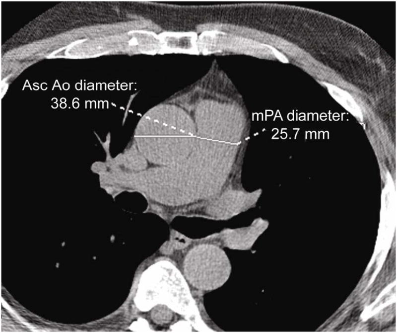
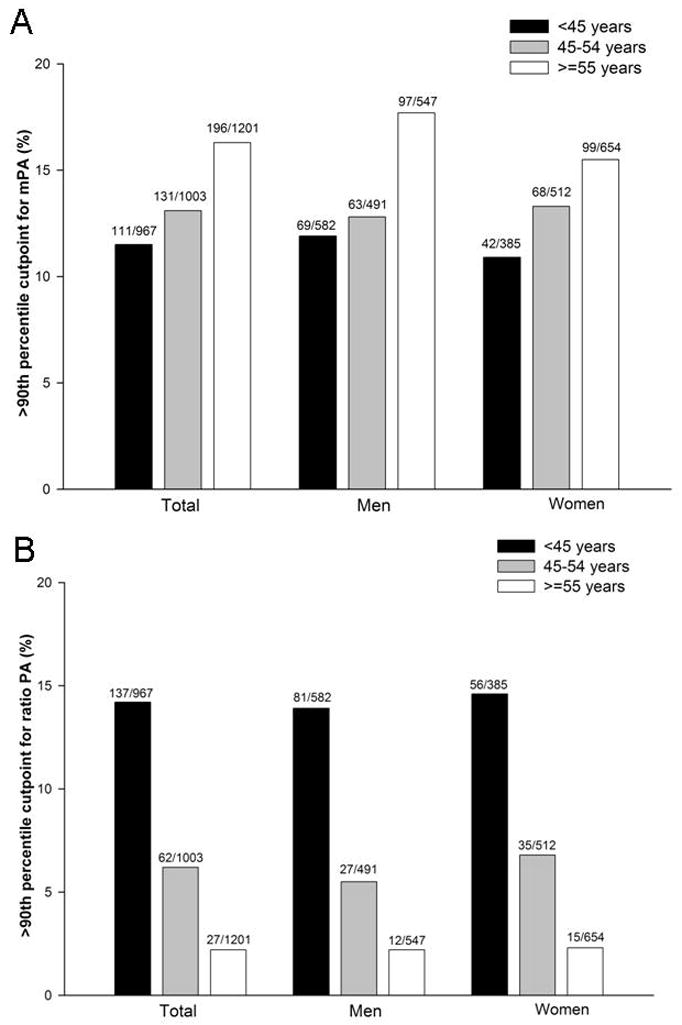
Methods and results: In 3171 men and women (mean age, 51±10 years; 51% men) from the Framingham Heart Study, a noncontrast, ECG-gated, 8-slice cardiac multidetector CT was performed. We measured the mPA and transverse axial diameter of the ascending aorta at the level of the bifurcation of the right pulmonary artery and calculated the ratio PA. We defined the healthy referent cohort (n=706) as those without obesity, hypertension, current and past smokers, chronic obstructive pulmonary disease, history of pulmonary embolism, diabetics, cardiovascular disease, and heart valve surgery. The mean mPA diameter in the overall cohort was 25.1±2.8 mm and mean ratio PA was 0.77±0.09. The sex-specific 90th percentile cutoff value for mPA diameter was 28.9 mm in men and 26.9 mm in women and was associated with increase risk for self-reported dyspnea (adjusted odds ratio, 1.31; P=0.02). The 90th percentile cutoff value for ratio PA of the healthy referent group was 0.91, similar between sexes but decreased with increasing age (range, 0.82-0.94), though not associated with dyspnea.
Conclusions: For simplicity, we established 29 mm in men and 27 mm in women as sex-specific normative reference values for mPA and 0.9 for ratio PA.
Figures
References
-
- Kuriyama K, Gamsu G, Stern RG, Cann CE, Herfkens RJ, Brundage BH. CT-determined pulmonary artery diameters in predicting pulmonary hypertension. Invest Radiol. 1984;19:16–22. - PubMed
-
- Haimovici JB, Trotman-Dickenson B, Halpern EF, Dec GW, Ginns LC, Shepard JA, McLoud TC. Relationship between pulmonary artery diameter at computed tomography and pulmonary artery pressures at right-sided heart catheterization. Massachusetts General Hospital Lung Transplantation Program. Acad Radiol. 1997;4:327–334. - PubMed
-
- Edwards PD, Bull RK, Coulden R. CT measurement of main pulmonary artery diameter. Br J Radiol. 1998;71:1018–1020. - PubMed
-
- Ng CS, Wells AU, Padley SP. A CT sign of chronic pulmonary arterial hypertension: the ratio of main pulmonary artery to aortic diameter. J Thorac Imaging. 1999;14:270–278. - PubMed
-
- Beiderlinden M, Kuehl H, Boes T, Peters J. Prevalence of pulmonary hypertension associated with severe acute respiratory distress syndrome: predictive value of computed tomography. Intensive Care Med. 2006;32:852–857. - PubMed
Publication types
MeSH terms
Grants and funding
LinkOut - more resources
Full Text Sources
Medical
Miscellaneous
