. 2011 Dec 18;14(1):87-92.
doi: 10.1038/ncb2395.

The adaptor protein CRK is a pro-apoptotic transducer of endoplasmic reticulum stress

Affiliations

The adaptor protein CRK is a pro-apoptotic transducer of endoplasmic reticulum stress

Kathryn Austgen et al. Nat Cell Biol. .

Abstract

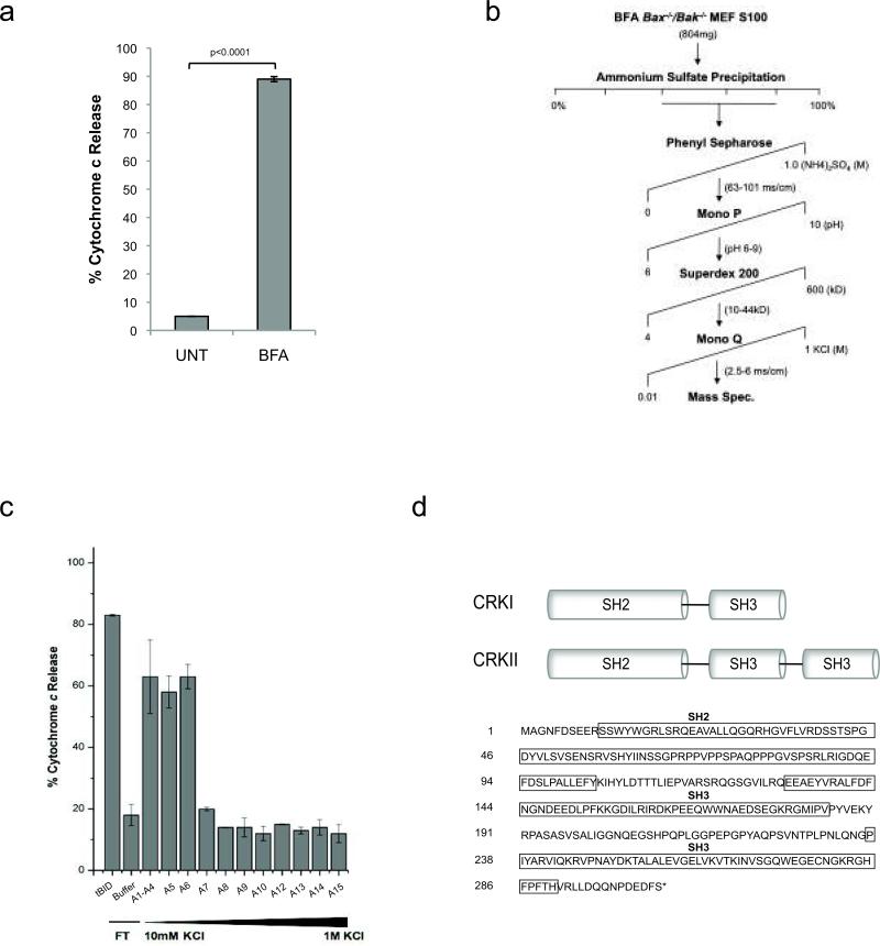
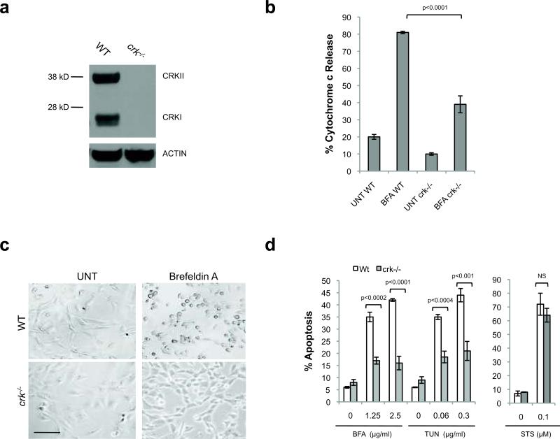
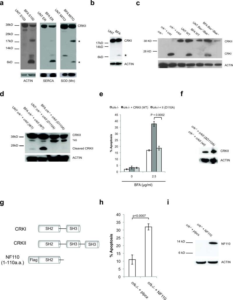
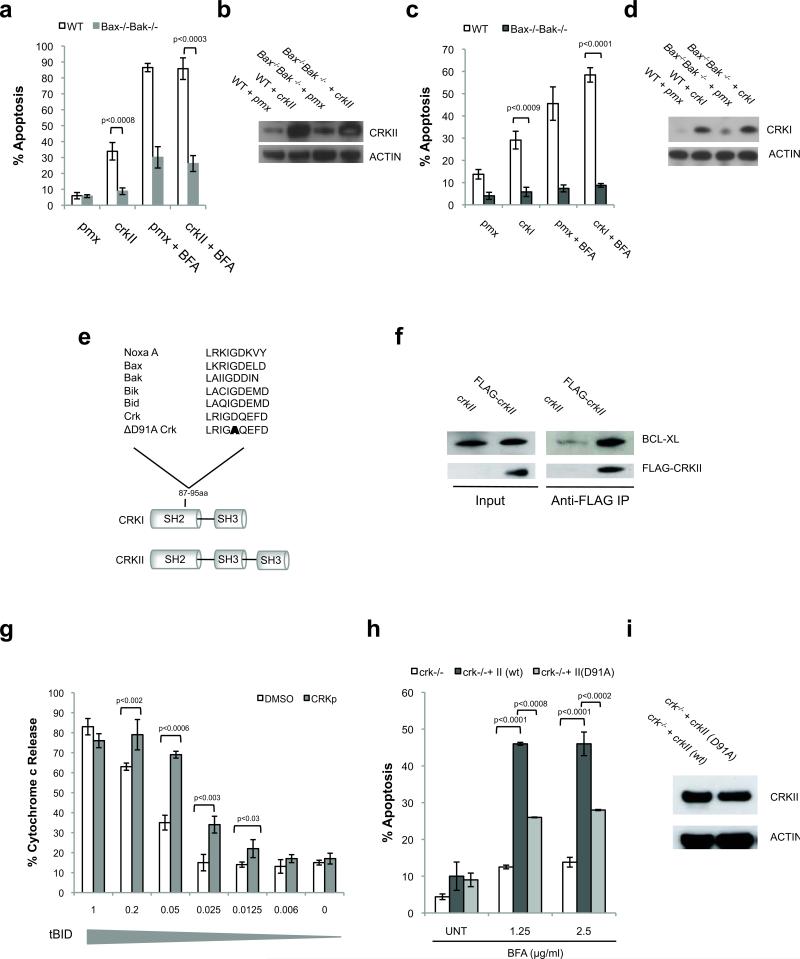
Excessive demands on the protein-folding capacity of the endoplasmic reticulum (ER) cause irremediable ER stress and contribute to cell loss in a number of cell degenerative diseases, including type 2 diabetes and neurodegeneration. The signals communicating catastrophic ER damage to the mitochondrial apoptotic machinery remain poorly understood. We used a biochemical approach to purify a cytosolic activity induced by ER stress that causes release of cytochrome c from isolated mitochondria. We discovered that the principal component of the purified pro-apoptotic activity is the proto-oncoprotein CRK (CT10-regulated kinase), an adaptor protein with no known catalytic activity. Crk(-/-) cells are strongly resistant to ER-stress-induced apoptosis. Moreover, CRK is cleaved in response to ER stress to generate an amino-terminal M(r)~14K fragment with greatly enhanced cytotoxic potential. We identified a putative BH3 (BCL2 homology 3) domain within this N-terminal CRK fragment, which sensitizes isolated mitochondria to cytochrome c release and when mutated significantly reduces the apoptotic activity of CRK in vivo. Together these results identify CRK as a pro-apoptotic protein that signals irremediable ER stress to the mitochondrial execution machinery.


Figures

Figure 1
Figure 1. Biochemical purification of ER stress apoptotic activity identifies proto-oncogene CT-10-regulated kinase (CRK)
(a) Induction of cytochrome c release from isolated Jurkat mitochondria by cytosolic extracts (S100) from untreated (UNT) and 24h Brefeldin A (BFA) 2.5 μg/ml treated Bax-/-Bak-/- MEFs. (b) FPLC purification scheme of cytochrome c releasing activity (CcRA) present in BFA Bax-/-Bak-/- S100. Active fractions from each purification step are indicated. (c) CcRA assay of the fractions from the final step of the purification (MonoQ ion exchange gradient). (d) Diagram of CRK isoforms, domains, and amino acid sequence.
Figure 2
Figure 2. Crk-/- MEFs are significantly resistant to ER stress-induced apoptosis
(a, b) 18h BFA (2.5 μg/ml)-treated crk-/- MEF S100 contains significantly less CcRA in comparison to 18h BFA-treated (2.5 μg/ml) wild-type MEF S100. n=3, error bars = sd. (c) crk-/- MEFs are visually resistant (phase contrast) to ER stress-induced apoptosis (BFA 2.5μg/ml). Scale bar, 100μm. (d) crk-/- MEFs are strongly resistant to BFA and TUN-induced apoptosis, but equally sensitive to staurosporine (STS), in comparison to wild-type MEFs. n=3, error bars = sd.
Figure 3
Figure 3. CRKI or CRKII restores sensitivity of crk-/- MEFs to ER stress-induced apoptosis
(a, b) Transient expression of CRKI or CRKII sensitizes crk-/- MEFs to BFA-induced apoptosis. n=3, error bars = sd. (c-e) Stable CRKII expression in crk-/- MEFs rescues sensitivity to 24h BFA- and 18h TUN-induced apoptosis, but does not change sensitivity to STS-induced apoptosis. n=3, error bars = sd. Scale bar, 100μm. (f, g) Stable overexpression of CRKII in wild-type MEFs further increases sensitivity to 18h BFA-induced apoptosis. n=3, error bars = sd. (h, i) Transient overexpression of CRKI sensitizes WT MEFs to 18h BFA (1.25 μg/ml)-induced apoptosis. n=3, error bars = sd.
Figure 4
Figure 4. CRK is proteolytically cleaved into an apoptotic signal upon irremediable ER stress
(a) Upon 24h BFA (2.5 μg/ml) treatment of Bax-/-Bak-/- MEFs, full-length CRKII is depleted in the cytosol and at the ER. CRKII-specific fragments (*) appear in the cytosol, ER, and mitochondria. (b) Transiently expressed CRKI is also cleaved upon 24h BFA (2.5 μg/ml) treatment in crk-/- MEFs. * = CRKI-specific fragment. (c) Loss of full-length, endogenous CRKI and CRKII observed upon 18h BFA treatment of WT and Bax-/-Bak-/- MEFs. (d) Upon ER stress, CRK is cleaved at D110. Mutation of this site (D110A) in CRKII prevents cleavage following 24h 2.5 μg/ml BFA treatment in stably reconstituted in crk-/- MEFs. (e, f) CrkII (D110A) is not able to rescue crk-/- MEF sensitivity to ER stress-induced apoptosis induced by 24h 2.5 μg/ml BFA, in contrast to crk-/- MEFs stably expressing wild-type (WT) CRKII. n=3, error bars = sd. (g) Diagram of CRK (1-110a.a.) cleavage fragment (NF110) produced upon ER stress. (h, i) Transient expression of NF110 induces apoptosis independent of ER stress. n=3, error bars = sd.
Figure 5
Figure 5. CRKII contains a putative BH3 domain and triggers BAX/BAK-dependent apoptosis
(a, b) CRKII and empty vector (pmx) were transiently overexpressed in WT and Bax-/-Bak-/- MEFs using retroviral infection. 24h post retroviral infection cells were treated with BFA (2.5 μg/ml) for an additional 24h and analyzed for Annexin-V expression by flow cytometry. n=3, error bars = sd. (c, d) CRKI and empty vector (pmx) were transiently overexpressed in WT and Bax-/-Bak-/- MEFs. 24h post transfection cells were treated an additional 18h with BFA (2.5 μg/ml) and analyzed for Annexin-V expression by flow cytometry. n=3, error bars = sd. (e) The sequences of the putative BH3-only domain of CRK and the “BH3 domain” point mutation D91A are aligned against BH3 domains of several known BH3-only proteins. (f) 293 cells were transiently transfected 24h with Flag-crkII or untagged crkII, then treated 14h BFA (1.25μg/ml). Lysates were incubated with FLAG-specific agarose beads. Beads were immunoblotted for endogenous BCL-XL. (g) Cytochrome c release from isolated Jurkat mitochondria incubated with decreasing doses of tBID and CRK BH3 domain peptide. n=3, error bars = sd. (h, i) Stable reconstitution of D91A crkII into crk-/- MEFs is significantly less effective at restoring ER stress-induced apoptosis (24h BFA treatment) in comparison to expression of wild-type crkII. n=3, error bars = sd.

References

    1. Marciniak SJ, Ron D. Endoplasmic reticulum stress signaling in disease. Physiol Rev. 2006;86:1133–1149. - PubMed
    1. Lin JH, Walter P, Yen TS. Endoplasmic reticulum stress in disease pathogenesis. Annu Rev Pathol. 2008;3:399–425. - PMC - PubMed
    1. Shore GC, Papa FR, Oakes SA. Signaling cell death from the endoplasmic reticulum stress response. Curr Opin Cell Biol. 2011;23:143–149. - PMC - PubMed
    1. Oakes SA, Lin SS, Bassik MC. The control of endoplasmic reticulum-initiated apoptosis by the BCL-2 family of proteins. Curr Mol Med. 2006;6:99–109. - PubMed
    1. Han D, et al. IRE1alpha kinase activation modes control alternate endoribonuclease outputs to determine divergent cell fates. Cell. 2009;138:562–575. - PMC - PubMed

Publication types

MeSH terms

Substances