. 2012 Apr;40(4):995-1004.
doi: 10.3892/ijo.2011.1297. Epub 2011 Dec 13.

Drug resistance to paclitaxel is not only associated with ABCB1 mRNA expression but also with drug accumulation in intracellular compartments in human lung cancer cell lines

Affiliations

Drug resistance to paclitaxel is not only associated with ABCB1 mRNA expression but also with drug accumulation in intracellular compartments in human lung cancer cell lines

Masanori Shimomura et al. Int J Oncol. 2012 Apr.

Abstract

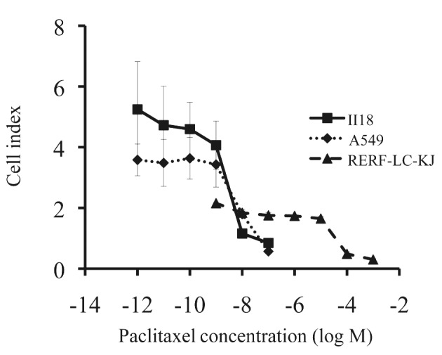
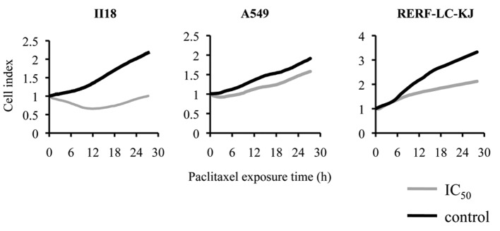
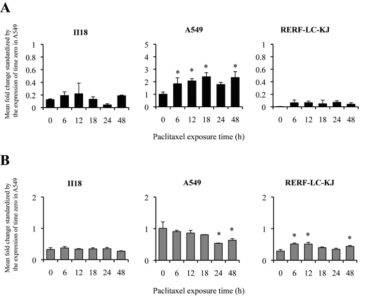
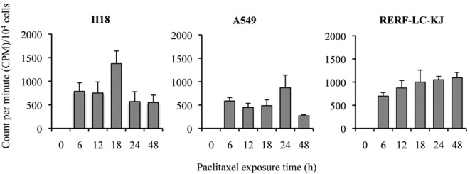
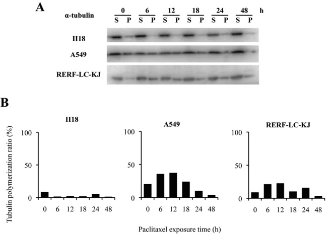
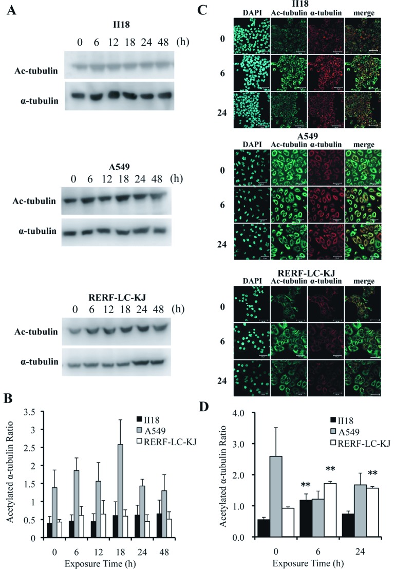
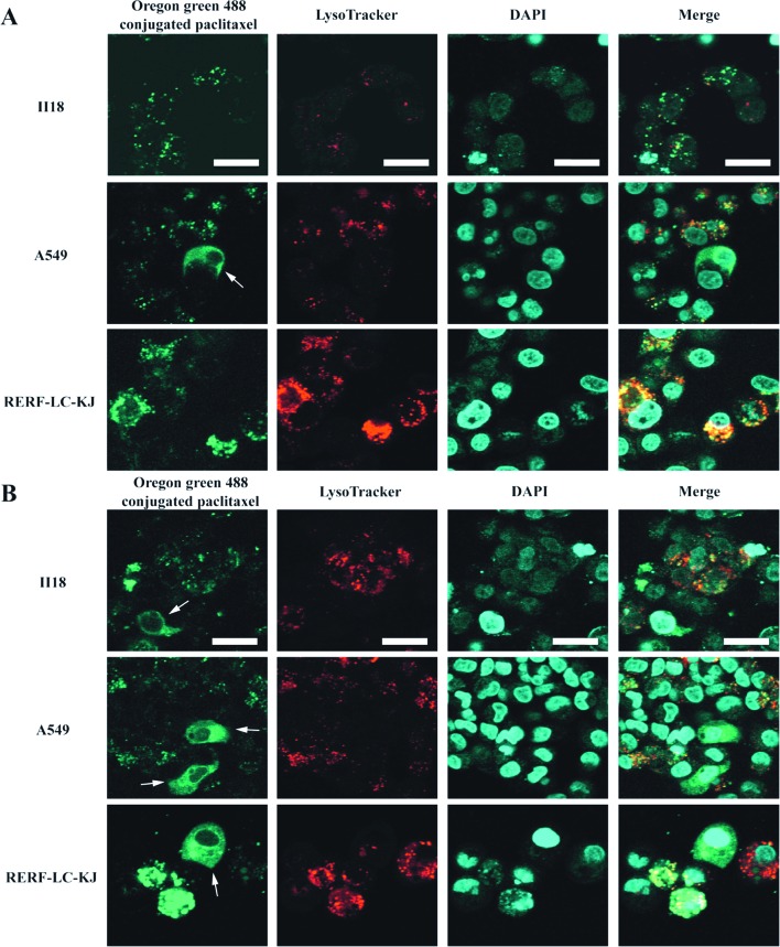
In order to clarify the mechanisms of resistance to paclitaxel in lung cancer, three human lung cancer cell lines which exhibit different sensitivity to paclitaxel were investigated from the following viewpoints: overexpression of ATP-binding cassette, sub-family B, member 1 (ABCB1), mutations on paclitaxel binding site of β-tubulin genes, quantity of polymerized tubulin and the intracellular localization of paclitaxel. ABCB1 expression was evaluated by real-time RT-PCR. No correlations were noted between the ABCB1 expression in the sensitive and resistant cell lines at the mRNA level. No mutations on the paclitaxel binding site of the β-tubulin genes were detected in either the resistant or sensitive cells. Live cell images obtained by confocal laser microscopy revealed that the resistant cell line, RERF-LC-KJ, had more accumulation of Oregon Green® 488 conjugated paclitaxel in the lysosomal and extra-lysosomal compartments of cytoplasm than other cell lines. The results obtained in this study indicated that the changes in the subcellular localization could contribute to the production of paclitaxel resistance in lung cancer cell lines. Further studies should be conducted to elucidate the molecular mechanisms that differentiate the intracellular localization of paclitaxel.


Figures

Figure 1
Figure 1
The effect of paclitaxel varied remarkably between the three lung cancer cell lines. The results of xCELLigence® system were used to measure the cell index, defined as the value reflecting the surface area covered by the living cells. Points, mean of one triplicated experiment; bars, SD.
Figure 2
Figure 2
The cytotoxicity of paclitaxel under concentrations required to inhibit growth by 50% (IC50) appeared most prominently within 24 h after the exposure, regardless of the cell line. The cellular proliferation was monitored by xCELLigence® system. After 24 h of incubation, the cells were exposed to paclitaxel.
Figure 3
Figure 3
The mRNA levels of ABCB1 (A) changed with time and differed in each lung cancer cell line when exposed to 3.2 nM paclitaxel. The changes in the mRNA level of ABCC1 (B) were modest in the II18 and RERF-LC-KJ cell lines, whereas the expression level significantly decreased at 24 and 48 h in the A549 cell line. The mRNA levels of ABCB1 and ABCC1 were analyzed by real-time RT-PCR and calculated by the ΔΔCT method. The data are presented relative to the level of ABCB1 and ABCC1 at time zero in the A549 cell line. The expression levels were normalized by the housekeeping gene GAPDH. Columns, the mean of one triplicated experiment; bars, SD; *p<0.05 relative to respective untreated (0 h) cells.
Figure 4
Figure 4
The intracellular accumulation of 3.2 nM [3H]-paclitaxel increases with time in the RERF-LC-KJ cell line, until it reached a peak between 12–24 h. The values were calculated as the CPM value divided by the cell number of each well. Columns, the mean of one triplicated experiment; bars, SD.
Figure 5
Figure 5
Paclitaxel does not stabilize the microtubules in the II18 cell line for 48 h, whereas it does in the other cell lines, with a peak between 6 or 12 h. (A), Immunoblots of proteins from lung cancer cell lines with antibodies against α-tubulin. The loading quantity was equal to 10 μg proteins from three 3.3 μg total proteins from the cell lines exposed to 3.2 nM paclitaxel. Cells were lysed in hypotonic buffer for 5 min at 37°C followed by separation of the soluble (S) and polymerized (P) fractions by centrifugation. (B), The percentage of polymerized tubulin was obtained by dividing the densitometric value of the polymerized tubulin (insoluble) by the total tubulin contents.
Figure 6
Figure 6
The expression of acetylated α-tubulin remained low for 48 h in the II18 cell line, whereas it increased with time in the RERF-LC-KJ cells. (A), The expressions of acetylated α-tubulin and α-tubulin were analyzed with the Western blot method using proteins extracted from cells exposed to 3.2 nM (−8.5 logM) paclitaxel for 6, 12, 18, 24 and 48 h and untreated (0 h). (B), The ratio of the expression level of acetylated α-tubulin to the total α-tubulin was measured. Columns, the mean of value of three experiments; bar, SD. (C), Cellular localization of acetylated microtubules (green) in cells exposed to paclitaxel for 6 and 24 h and untreated cells. The cells were stained with acetylated α-tubulin antibody, microtubules (red) with α-tubulin antibody and nuclei (blue) with DAPI. Bar, 50 μm. (D), The ratio of the expression of acetylated α-tubulin to that of α-tubulin obtained with immunofluorescent staining was calculated from integrating each fluorescent intensity on a series of stacked images with a thickness of 1 μm. Columns, mean of value obtained from 3 microscopic fields; bar, SD; **p<0.01 relative to respective untreated (0 h) cells.
Figure 7
Figure 7
Oregon Green® 488-conjugated paclitaxel made it possible to visualize not only the normal microtubules formation, but also the aggregated vesicle formation in some of the RERF-LC-KJ cells, whereas in the other cell lines these vesicular formations were not remarkable. (A and B), Representative confocal images of lung cancer cell lines after the incubation of Oregon Green® 488 conjugated paclitaxel for 6 and 24 h, respectively, followed by staining with 100 nM LysoTracker® DND-99 (red) and DAPI (blue). Cellular microtubules are highlighted by white arrows. Bars, 20 μm.

References

    1. Sawabata N, Asamura H, Goya T, et al. Japanese Lung Cancer Registry Study: first prospective enrollment of a large number of surgical and nonsurgical cases in 2002. J Thorac Oncol. 2010;5:1369–1375. - PubMed
    1. Pujol JL, Barlesi F, Daures JP. Should chemotherapy combinations for advanced non-small cell lung cancer be platinum-based? A meta-analysis of phase III randomized trials. Lung Cancer. 2006;51:335–345. - PubMed
    1. Ambudkar SV, Kimchi-Sarfaty C, Sauna ZE, Gottesman MM. P-glycoprotein: from genomics to mechanism. Oncogene. 2003;22:7468–7485. - PubMed
    1. Huang Y, Sadee W. Membrane transporters and channels in chemoresistance and sensitivity of tumor cells. Cancer Lett. 2006;239:168–182. - PubMed
    1. Lee JT, Jr, Steelman LS, McCubrey JA. Phosphatidylinositol 3′-kinase activation leads to multidrug resistance protein-1 expression and subsequent chemoresistance in advanced prostate cancer cells. Cancer Res. 2004;64:8397–8404. - PubMed

MeSH terms

Substances