. 2011 Dec 18;7(3):174-9.
doi: 10.1038/nnano.2011.223.

Intracellular recordings of action potentials by an extracellular nanoscale field-effect transistor

Affiliations

Intracellular recordings of action potentials by an extracellular nanoscale field-effect transistor

Xiaojie Duan et al. Nat Nanotechnol. .

Abstract

The ability to make electrical measurements inside cells has led to many important advances in electrophysiology. The patch clamp technique, in which a glass micropipette filled with electrolyte is inserted into a cell, offers both high signal-to-noise ratio and temporal resolution. Ideally, the micropipette should be as small as possible to increase the spatial resolution and reduce the invasiveness of the measurement, but the overall performance of the technique depends on the impedance of the interface between the micropipette and the cell interior, which limits how small the micropipette can be. Techniques that involve inserting metal or carbon microelectrodes into cells are subject to similar constraints. Field-effect transistors (FETs) can also record electric potentials inside cells, and because their performance does not depend on impedance, they can be made much smaller than micropipettes and microelectrodes. Moreover, FET arrays are better suited for multiplexed measurements. Previously, we have demonstrated FET-based intracellular recording with kinked nanowire structures, but the kink configuration and device design places limits on the probe size and the potential for multiplexing. Here, we report a new approach in which a SiO2 nanotube is synthetically integrated on top of a nanoscale FET. This nanotube penetrates the cell membrane, bringing the cell cytosol into contact with the FET, which is then able to record the intracellular transmembrane potential. Simulations show that the bandwidth of this branched intracellular nanotube FET (BIT-FET) is high enough for it to record fast action potentials even when the nanotube diameter is decreased to 3 nm, a length scale well below that accessible with other methods. Studies of cardiomyocyte cells demonstrate that when phospholipid-modified BIT-FETs are brought close to cells, the nanotubes can spontaneously penetrate the cell membrane to allow the full-amplitude intracellular action potential to be recorded, thus showing that a stable and tight seal forms between the nanotube and cell membrane. We also show that multiple BIT-FETs can record multiplexed intracellular signals from both single cells and networks of cells.


Conflict of interest statement

Competing financial interests: The authors declare that they have no competing financial interests.

Figures

Figure 1
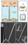
Figure 1. Schematics and structural characterization of the BIT-FET
a, schematics showing (left) a cell coupled to a BIT-FET and (right) the variation in device conductance, (G), during an action potential, where S, D, Vm, and t correspond to source electrode, drain electrode, membrane potential, and time, respectively. The SiO2 nanotube connects the cytosol (orange) to the p-type SiNW FET, and together with the SiO2 passivation excludes the extracellular medium (light blue) from the active device channel. The schematic structures on the membrane represent different ion channels, and are not scaled to the true size of the BIT-FET. b, SEM image of a GeNW branch on a SiNW with an orientation near the surface normal. Inset, SEM image of an Au nanodot on a SiNW prior to GeNW growth. c, SEM image of a GeNW/SiNW heterostructure coated with ALD SiO2. Magnified images of the top and bottom are shown in Fig. S2. d, SEM image of a final nanotube on a SiNW. Insets, magnified images of the top and bottom of the nanotube. Scale bars, 100 nm in inset of b, 200 nm in all others.
Figure 2
Figure 2. Water gate characterization and bandwidth analysis
a, SEM image of a BIT-FET device (S-D1) and control device (S-D2). b and c, Water gate, Vwg, responses prior to and after GeNW etching, respectively. Blue, (S-D1); red, (S-D2). d, pulsed Vwg with 0.1 ms rise/fall time, 1 ms duration and 100 mV amplitude (upper), and the corresponding conductance change of a BIT-FET device (black, lower). The red trace is the pure field-effect response after removing the capacitive signals of the passivated metal electrodes (see Supplementary Methods). e, baseline to plateau conductance change of the same BIT-FET device as in d versus pulse rise/fall time. The change was measured as an average over data 0.2-0.5 ms after the start of the pulse. Pulse amplitude was kept at 100 mV, and duration was ten times the rise/fall time in all measurements. f, Calculated bandwidth of the BIT-FET device versus nanotube inner diameter (ALD SiO2 thickness was the same as the nanotube inner diameter, and the nanotube length was fixed at 1.5 μm). The black and red symbols correspond to upper and lower limits, respectively (see Supplementary Methods). Inset, calculated change of the potential at the SiNW FET surface Vn (normalized with the step change V0 of potential at the nanotube opening) versus time.
Figure 3
Figure 3. Intracellular action potential recording
a, a representative trace reflects the transition from extra- to intra- cellular recording. b, magnified trace of the part in the black dashed rectangle in a. c, magnified trace of the peak in the blue dashed rectangle in b. The stars in b and c mark the position of extracellular spikes. d, magnified trace of the peak in red dashed rectangle in a. e, the trace corresponding to the second entry of the nanotube around the same position on the cell. The potential was calibrated using the sensitivity values measured on phospholipid-modified devices by quasi-static Vwg measurement (e.g. blue trace in Fig. 2c) and pulsed Vwg measurement with 0.1 ms pulse rise/fall time (same for Fig. 4). The sensitivity obtained from these two measurements is same as discussed before.
Figure 4
Figure 4. Multiplexed intracellular recording
a, Differential interference contrast microscopy (DIC) image of two BIT-FET devices (position marked with dots) coupled to a single cardiomyocyte cell, with cell boundary marked by the yellow dashed line. b, The simultaneously recorded traces from the two devices in a, corresponding to the transition from extra- to intra- cellular signal. The transition happened in sequential manner. The break mark labels the ~1 s discontinuity between the two adjacent traces. c, representative trace of stable intracellular action potentials recorded ~120 s after the internalization of the devices in a. d, DIC image of three BIT-FET devices coupled to a layer of beating cardiomycyte cell network (from a different PDMS/cell sample than in a). e, representative traces recorded simultaneously from the devices shown in d. The three devices exhibit intracellular action potential signals from different cells in the cell network. We note that devices used in a and d have different sensitivities (and are also different from the one used in Fig. 3). These differences are primarily due to variations in Ge over-coating during GeNW growth (see Supplementary Information). The potential was calibrated using the sensitivity values measured for each individual device, and all devices yield corresponding intracellular action potential values with full amplitude of 75-100 mV (independent of this conductance/sensitivity variation).

Comment in

References

    1. Sakmann B, Neher E. Patch clamp techniques for studying ionic channels in excitable membranes. Ann Rev Physiol. 1984;46:455–472. - PubMed
    1. Molleman A. Patch clamping: an introductory guide to patch clamp electrophysiology. Wiley; Chichester, England: 2003.
    1. Rutten WLC. Selective electrical interfaces with the nervous system. Annu Rev Biomed Eng. 2002;4:407–452. - PubMed
    1. Purves RD. Microelectrode methods for intracellular recording and ionophoresis. Academic Press; London: 1981.
    1. Chorev E, Epsztein J, Houweling AR, Lee AK, Brecht M. Electrophysiological recordings from behaving animals-going beyond spikes. Curr Opin Neurobiol. 2009;19:513–519. - PubMed

Publication types