. 2012 Feb 10;287(7):4982-95.
doi: 10.1074/jbc.M111.306860. Epub 2011 Dec 16.

Identification and functional impact of homo-oligomers of the human proton-coupled folate transporter

Affiliations

Identification and functional impact of homo-oligomers of the human proton-coupled folate transporter

Zhanjun Hou et al. J Biol Chem. .

Abstract

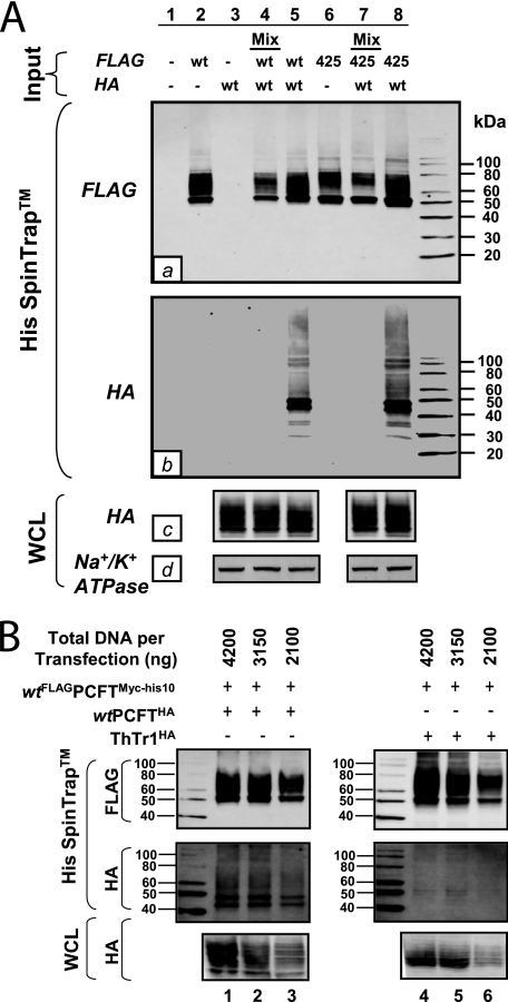
The proton-coupled folate transporter (PCFT; SLC46A1) is a proton-folate symporter that is abundantly expressed in solid tumors and normal tissues, such as duodenum. The acidic pH optimum for PCFT is relevant to intestinal absorption of folates and could afford a means of selectively targeting tumors with novel cytotoxic antifolates. PCFT is a member of the major facilitator superfamily of transporters. Because major facilitator superfamily members exist as homo-oligomers, we tested this for PCFT because such structures could be significant to PCFT mechanism and regulation. By transiently expressing PCFT in reduced folate carrier- and PCFT-null HeLa (R1-11) cells and chemical cross-linking with 1,1-methanediyl bismethanethiosulfonate and Western blotting, PCFT species with molecular masses approximating those of the PCFT dimer and higher order oligomers were detected. Blue native polyacrylamide gel electrophoresis identified PCFT dimer, trimer, and tetramer forms. PCFT monomers with hemagglutinin and His(10) epitope tags were co-expressed in R1-11 cells, solubilized, and bound to nickel affinity columns, establishing their physical associations. Co-expressing YPet and ECFP*-tagged PCFT monomers enabled transport and fluorescence resonance energy transfer in plasma membranes of R1-11 cells. Combined wild-type (WT) and inactive mutant P425R PCFTs were targeted to the cell surface by surface biotinylation/Western blots and confocal microscopy and functionally exhibited a "dominant-positive" phenotype, implying positive cooperativity between PCFT monomers and functional rescue of mutant by WT PCFT. Our results demonstrate the existence of PCFT homo-oligomers and imply their functional and regulatory impact. Better understanding of these higher order PCFT structures may lead to therapeutic applications related to folate uptake in hereditary folate malabsorption, and delivery of PCFT-targeted chemotherapy drugs for cancer.


Figures

FIGURE 1.
FIGURE 1.
hPCFT topology model. A topology model for hPCFT is shown, including 12 TMDs and internal N and C termini. The seven Cys residues are shown as black circles. Structurally and functionally important residues, as described in the “Introduction,” are labeled as black squares. The DXXGRR motif (positions 109–114) is shown as gray circles. Pro-425, for which its Arg mutant was used in current study, is shown as a black triangle. N-Glycosylation occurs at Asn-58 and Asn-68 (also shown as black circles).
FIGURE 2.
FIGURE 2.
Detection of putative hPCFT oligomers with a MTS homobifunctional cross-linker. The dghPCFTMyc-His6 construct was transiently expressed in hRFC- and hPCFT-null HeLa R1-11 cells. Cells were treated with the MTS-1-MTS bifunctional cross-linker at 4 °C for 30 min. Following cross-linking, plasma membranes were prepared, and membrane proteins were analyzed by a 4–20% Novex® Tris/glycine gel under non-reducing conditions and Western blotting with hPCFT-specific polyclonal antibody. In the absence of DTT treatment, unique bands (b and c (lane 3)) were identified that were not seen in the absence of cross-linker (lanes 1 and 2). Treatment of cross-linked proteins with DTT prior to running the gradient gel reversed cross-linking for bands b and c (lanes 4–10). In the figure, a ∼60-kDa protein was detected with hPCFT-specific antibody in cross-linked samples treated with DTT. This band was also detected in non-cross-linked samples treated with reducing agents (e.g. DTT or 2-mercaptoethanol). Although species a–c were all detected when the blot shown was probed with cMyc antibody (not shown), the ∼60-kDa species was not. NS, nonspecific.
FIGURE 3.
FIGURE 3.
Demonstration of putative hPCFT oligomers on blue native polyacrylamide gels. dgFLAGhPCFTMyc-His10 was transfected into R1-11 cells, and cells were harvested 48 h later. Plasma membranes were prepared for BN-PAGE, as described under “Experimental Procedures.” The sample and protein standards were run on a 4–16% BisTris gel and then transferred to a PVDF membrane. The protein standard lane was cut out, and the membrane was stained separately with CB G250. The rest of the blot was processed for Western analysis and developed with anti-FLAG (primary) and IRDye800-conjugated (secondary) antibodies. Immunoreactive proteins were detected on an Odyssey infrared imager. The figure shows putative tetrameric ((hPCFT)4), trimeric ((hPCFT)3), and dimeric ((hPCFT)2) species, as described under “Results.”
FIGURE 4.
FIGURE 4.
Co-transfections of WT and P425R hPCFTs. wtFLAGhPCFTMyc-His10, wthPCFTHA, and FLAGP425RMyc-His10 were transiently transfected into HeLa R1-11 cells either singly or in combination. Transfection efficiencies were monitored with pGL4.74[hRluc/TK] vector, which encodes Renilla luciferase under control of a thymidine kinase promoter, and luciferase assays. A, cells were assayed for transport at pH 5.5 with [3H]Mtx (0.5 μm) for 2 min at 37 °C. Transport results are normalized to Renilla luciferase activities; results are expressed relative to those for wthPCFTHA and are reported as mean values ± S.E. (error bars) from seven independent experiments. Compared with wthPCFTHA, the increases of relative transport of both dual transfectants (wthPCFTHA plus wtFLAGhPCFTMyc-His10 and wthPCFTHA plus FLAGP425RMyc-His10) are significantly higher (p < 0.01; paired t test). B, results are shown for surface hPCFT proteins labeled with sulfo-NHS-SS-biotin (0.25 mg/ml) and isolated on immobilized NeutrAvidinTM gel. Samples were analyzed by SDS-PAGE and Western blotting with His6- and HA-specific antibodies. Na+/K+ ATPase was used as a loading control. The molecular mass markers for SDS-PAGE are noted. NS, nonspecific.
FIGURE 5.
FIGURE 5.
Immunofluorescence staining of HA- and FLAG-tagged WT and mutant hPCFTs. A and B, results are shown for HeLa R1-11 cells transfected with wtFLAGhPCFTMyc-His10 (panels a, b, and d), wthPCFTHA (panels c, d, e, h, i, and j), and FLAGP425RMyc-His10 (panels f and g), either singly or together. Cells were fixed with 3.3% paraformaldehyde, permeabilized with 0.1% Triton X-100, and stained with Alexa Fluor® 488-conjugated (for FLAG-tagged proteins; green) or Alexa Fluor® 568-conjugated (for HA-tagged hPCFT; red) secondary antibodies. Slides were visualized with a Zeiss LSM-510 META NLO using a 63× water immersion lens. Staining is shown for the individual fluors and as a merged image.
FIGURE 6.
FIGURE 6.
Co-association of hPCFT monomers. R1-11 cells were co-transfected with wtFLAGhPCFTMyc-His10 or FLAGP425RMyc-His10, together with wthPCFTHA in fixed DNA amounts (4200 ng of DNA/sample) (A) or were co-transfected with wtFLAGhPCFTMyc-His10 combined with ThTr1HA or wthPCFTHA with different DNA amounts (4200, 3150, and 2100 ng/sample) (B) as 1:1 ratios for both constructs. Transfected cells were harvested, sonicated, and solubilized (whole cell lysate (WCL)), and the homogenates were fractionated on His SpinTrapTM columns, as detailed under “Experimental Procedures.” The whole cell lysates and the nickel column eluate fractions were separated by 4–20% Tris/glycine gels, followed by Western blotting with FLAG (labeled a in panel A) or HA (labeled b and c in panel A) antibodies. Loading for whole cell lysates was normalized by probing with antibody to Na+/K+ ATPase (labeled d in panel D).
FIGURE 7.
FIGURE 7.
Sensitized FRET emission between hPCFT monomers fused with mYPet and mECFP*. A, schematic of the mYPet and mECFP* tandem construct and their fusion constructs with hPCFT is shown in the upper panel. In the lower panel, results for determination of expression levels of individual mYPet and mECFP* proteins and the mYPet-mECFP* fusion protein by Western blotting are shown. Total cell lysates were prepared from non-transfected R5 HeLa cells and cells transiently transfected with mYPet, mECFP*, or mYPet-mECFP* constructs. Proteins were analyzed by SDS-PAGE and Western blots with detection using the Odyssey infrared imaging system and GFP (primary, mouse polyclonal; Abcam) and IRDye800-conjugated (secondary) antibodies. B, mYPet and mECFP* hPCFT fusion proteins were characterized for expression and transport function. Plasma membranes were prepared from non- and mock-pCDNA3 R1-11 cells and R1-11 cells transiently transfected with individual mYPet-hPCFT, hPCFT-mECFP*, and hPCFT-mYPet constructs. Plasma membrane preparations were isolated and separated by SDS-PAGE, followed by Western blot analysis and detection with the Odyssey infrared imaging system, using hPCFT-specific (top) and GFP-specific (middle) antibodies and IRDye800-conjugated secondary antibody. Transfected cells were also assayed for membrane transport (bottom). [3H]Mtx (0.5 μm) uptakes were measured for 2 min at 37 °C. Representative transport results are shown. C, for FRET assays, mYPet plus mECFP* (negative control), mYPet-mECFP* tandem (positive control), mYPet-hPCFT plus hPCFT-mECFP*, and hPCFT-mYPet plus hPCFT-mECFP* constructs were transiently transfected into R1-11 cells. R1-11 cells were also singly transfected with mYPet, mECFP*, mYPet-hPCFT, hPCFT-mECFP*, and hPCFT-mYPet constructs as controls (not shown). Forty-eight hours post-transfection, mYPet and mECFP* images were acquired on a confocal microscope, and total donor (mECFP*), total acceptor (mYPet), and sensitized FRET emissions (NET FRET) were calculated, as described under “Experimental Procedures.” FRET efficiency values from 15 separate cells (for each experimental sample and controls, as well) were used for statistical analysis. FRET efficiencies for both hPCFT dual transfections (mYPet-hPCFT plus hPCFT-mECFP* and hPCFT-mYPet plus hPCFT-mECFP*) were significantly higher than that for the negative control (mYPet plus mECFP*), with p < 0.0001 (***) and p = 0.0007 (**) (paired t test), respectively. For the co-transfections, the FRET efficiency of mYPet-hPCFT plus hPCFT-mECFP* is significantly higher than that of hPCFT-mYPet plus hPCFT-mECFP* (p = 0.0003; paired t test). D, images collected in the FRET channel were calculated, as described under “Experimental Procedures.” Representative images for each sample are shown. Calculated FRET values for both test samples (mYPet-hPCFT plus hPCFT-mECFP* and hPCFT-mYPet plus hPCFT-mECFP*) and controls (mYPet plus mECFP*, mYPet-mECFP*) are represented using a pseudocolor scale. NET FRET signals for both hPCFT samples were found mainly in the cell surface membranes, whereas significant FRET was observed intracellularly for the mYPet-mECFP* positive control. Error bars, S.E.
FIGURE 8.
FIGURE 8.
Dominant-positive interactions between WT and inactive P425R hPCFT. wtPCFTHA and FLAGP425RHA-Myc-His10 constructs were transiently transfected into R1-11 cells. The cells were assayed for transport with [3H]Mtx (0.5 μm) over 2 min at pH 5.5 and at 37 °C. Surface hPCFT proteins were labeled with sulfo-NHS-SS-biotin. The biotinylated proteins were isolated, deglycosylated, and analyzed by 4–20% Tris/glycine gels and Western blotting with HA-specific antibody, as described under “Experimental Procedures.” Relative levels of WT and P425R proteins were measured by densitometry. A shows the linear (dashed) line predicted for functionally non-interacting hPCFT monomers, as described under “Results.” The data shown are for transport activities in excess of the low residual level for P425R plotted against the fraction of WT hPCFT in the WT/P425R hPCFT mixtures, as calculated from densitometry measurements from three independent experiments. Results are mean values ± S.E. (error bars). Statistics and correlation coefficients were calculated by Prism software (version 4.0). A clear non-linear dose response is observed (solid line). B shows a representative Western blot of deglycosylated wtPCFTHA and FLAGP425RHA-Myc-His10 proteins. The calculated fractions of WT hPCFT to total hPCFT proteins from each sample are noted below each lane with S.E. values from three independent experiments in parentheses. Experimental details are provided under “Experimental Procedures”.
FIGURE 9.
FIGURE 9.
Proposed reaction scheme for hPCFT-mediated cellular uptake involving cooperative interactions between hPCFT monomers. Based on the “alternate access model” for secondary transporters, such as Lac Y (60), adapted from that of Unal et al. (30) for monomeric PCFT, an analogous reaction scheme is depicted for hPCFT-mediated transport that incorporates the functional impact of hPCFT oligomerization. The model starts from the outward facing unloaded dimer, followed by the ordered binding of the co-transported protons (step 1) and (anti)folate substrates (step 2), which triggers a conformational change resulting in simultaneous transition of the two hPCFT monomers to an inward facing state (step 3). This is followed by an ordered release of substrates (step 4) and then protons (step 5) into the cytoplasm. The unloaded homo-oligomeric unit then returns to the outward facing state (step 6) to complete the transport cycle. In this model, the two hPCFT monomers are suggested to function cooperatively in facilitating substrate and proton binding, conformational changes, and substrate and proton release. For further details, see “Discussion.”

Similar articles

Cited by

References

    1. Stokstad E. L. R. (ed) (1990) Historical Perspective on Key Advances in the Biochemistry and Physiology of Folates, pp. 1–21, Wiley-Liss, New York
    1. Zhao R., Matherly L. H., Goldman I. D. (2009) Membrane transporters and folate homeostasis. Intestinal absorption and transport into systemic compartments and tissues. Expert. Rev. Mol. Med. 11, e4. - PMC - PubMed
    1. Zhao R., Goldman I. D. (2007) The molecular identity and characterization of a proton-coupled folate transporter–PCFT. Biological ramifications and impact on the activity of pemetrexed. Cancer Metastasis Rev. 26, 129–139 - PubMed
    1. Nakai Y., Inoue K., Abe N., Hatakeyama M., Ohta K. Y., Otagiri M., Hayashi Y., Yuasa H. (2007) Functional characterization of human proton-coupled folate transporter/heme carrier protein 1 heterologously expressed in mammalian cells as a folate transporter. J. Pharmacol. Exp. Ther. 322, 469–476 - PubMed
    1. Qiu A., Jansen M., Sakaris A., Min S. H., Chattopadhyay S., Tsai E., Sandoval C., Zhao R., Akabas M. H., Goldman I. D. (2006) Identification of an intestinal folate transporter and the molecular basis for hereditary folate malabsorption. Cell 127, 917–928 - PubMed

Publication types

MeSH terms