. 2012 Feb 15;31(4):842-55.
doi: 10.1038/emboj.2011.465. Epub 2011 Dec 16.

Identification and characterization of a resident vascular stem/progenitor cell population in preexisting blood vessels

Affiliations

Identification and characterization of a resident vascular stem/progenitor cell population in preexisting blood vessels

Hisamichi Naito et al. EMBO J. .

Abstract

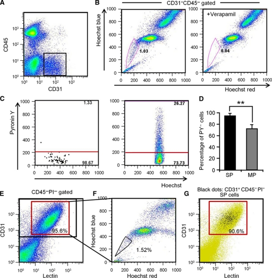
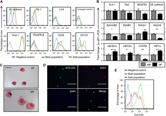
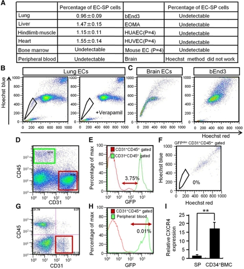
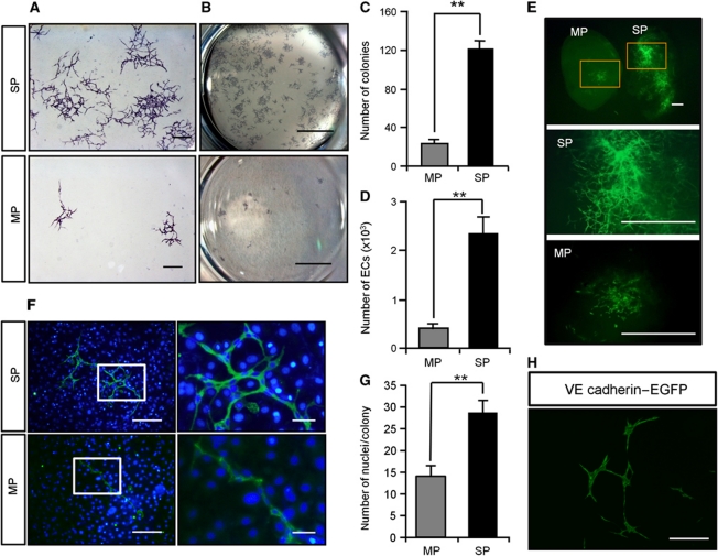
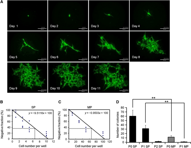
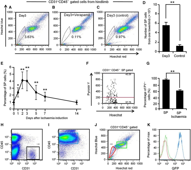
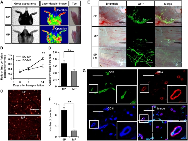
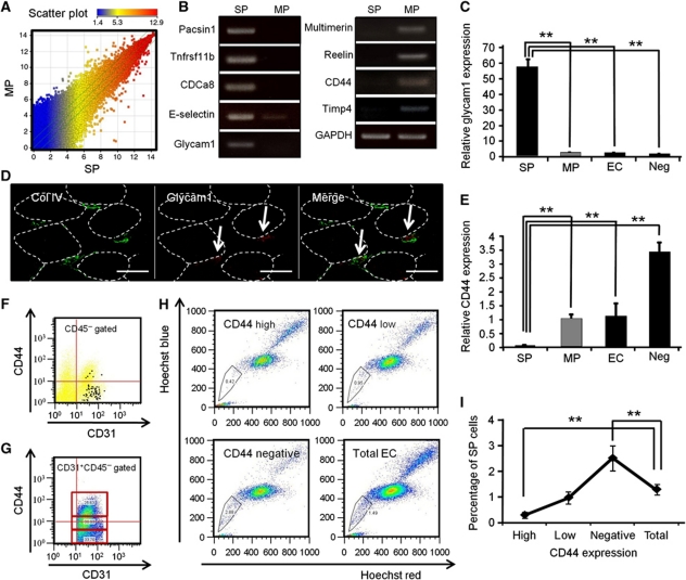
Vasculogenesis, the in-situ assembly of angioblast or endothelial progenitor cells (EPCs), may persist into adult life, contributing to new blood vessel formation. However, EPCs are scattered throughout newly developed blood vessels and cannot be solely responsible for vascularization. Here, we identify an endothelial progenitor/stem-like population located at the inner surface of preexisting blood vessels using the Hoechst method in which stem cell populations are identified as side populations. This population is dormant in the steady state but possesses colony-forming ability, produces large numbers of endothelial cells (ECs) and when transplanted into ischaemic lesions, restores blood flow completely and reconstitutes de-novo long-term surviving blood vessels. Moreover, although surface markers of this population are very similar to conventional ECs, and they reside in the capillary endothelium sub-population, the gene expression profile is completely different. Our results suggest that this heterogeneity of stem-like ECs will lead to the identification of new targets for vascular regeneration therapy.


Conflict of interest statement

The authors declare that they have no conflict of interest.

Figures

Figure 1
Figure 1
Identification of endothelial side population cells. (A) Flow cytometric analysis of hind limb ECs from wild-type mice. (B) Hoechst 33342 staining of CD31+CD45 ECs gated as shown in (A). Note that verapamil selectively prevents Hoechst exclusion from EC-SP cells. (C) Incorporation of Pyronin Y (PY) in EC-SP (left-hand side) and EC-MP (right-hand side) cells. (D) Quantitative evaluation of PY cells among EC-SP (SP) and EC-MP (MP) cells. Error bars are ±s.e.m. **P<0.01 (n=7). (E) Flow cytometric analysis of mouse hind limb ECs after in-vivo infusion of lectin. Lectin-positive cells among the CD31+CD45 cells are shown in the red gate and total CD31+CD45 cells are shown in the black gate. 95.9±0.2% (n=6) of the CD31+CD45 ECs were lectin positive. (F) Hoechst staining of lectin+ CD31+CD45 cells. (G) Representative flow cytometric plots of EC-SP cells (black dots). The lectin-positive population is shown in the red gate. 90.6±1.4% (n=4) of the EC-SP cells were lectin positive.
Figure 2
Figure 2
Characterization of endothelial side population cells. (A) Histogram showing expression levels of surface markers in EC-SP, EC-MP cells and the negative control. (B) Quantitative RT–PCR analysis of mRNA as indicated in EC-SP and EC-MP cells, corrected for expression of the control gene GAPDH. Of the endothelial genes, Notch4 was significantly lower in EC-SP cells. Expression levels of the ABC transporter ABCG2 and ABCB1a (MDR1a) were higher in EC-SP cells. Expression of chemokine receptor CXCR4 and hypoxia-inducible factor (HIF1a) was higher in EC-SP cells. Error bars are ±s.e.m. **P<0.01, *P<0.05 (n>6). (C) Haematoxylin and eosin staining of EC-SP and EC-MP cells isolated by FACS. (D) Freshly isolated cells from hind limb were stained with Ac-LDL and then Hoechst staining was performed to detect EC-SP cells. EC-SP cells were cytospun onto slides and Ac-LDL uptake was evaluated. Some of the EC-SP cells showed weak uptake of Ac-LDL, but all were positive. (E) Intensity of Ac-LDL uptake was evaluated by FACS analysis. As indicated, Ac-LDL uptake was observed in EC-SP cells but was lower than in EC-MP cells. Scale bars, 10 μm (C) and 100 μm (D).
Figure 3
Figure 3
EC-SP cells are present in different organs and are not derived from BM in BM chimeric mice. (A) The ECs from several organs and cultured cell lines as indicated were stained with Hoechst. Percentages of the EC-SP cells are shown in the table. There were few CD31+CD45 ECs in the bone marrow and peripheral blood; SP cells were hardly detected at all. In the EC lines (HUVEC, HUAEC, bEnd3, and EOMA), SP cells were not detected. Of note, the EC-SP phenotype disappeared after culturing primary ECs from hind limbs. (B) One example showing EC-SP cells of lung that disappeared following verapamil treatment. (C) In the brain, a stereotypic EC-SP pattern is not observed and there are no EC-SP cells within the bEnd3 population. (DF) BM cells from GFP mice were transplanted into lethally irradiated wild-type mice. Four weeks after transplantation, cells from hind limbs were analysed. (D) Representative flow cytometric plots of cells from hind limb muscle. CD31+CD45 EC fraction (red) and CD31CD45+ peripheral blood fraction (green) are gated. (E) Histogram of CD31+CD45 ECs and CD31CD45+ peripheral blood cells obtained from hind limbs. Almost all blood cells (green line) after transplantation were GFP positive. Approximately 4% of CD31+CD45 ECs (red line) were weakly GFP positive (GFPdim). GFPdim EC population is shown in arrowed region. (F) Hoechst staining of GFPdim ECs. The SP phenotype was not seen. (G, H) Analysis of hind limb muscle cells from newborn transplantation model. (G) Representative flow cytometric plots of cells from hind limb muscle of BM chimeric mice; CD31+CD45 EC fraction is gated (red). (H) Histogram showing GFP intensity of CD31+CD45 ECs obtained from hind limb and peripheral blood. In this model, GFP-positive CD31+CD45 ECs make up <0.01% of total CD31+CD45 ECs, suggesting no major contribution of BM cells to EC-SP cells. (I) Quantitative evaluation of CXCR4 mRNA expression in EC-SP cells and CD34+ bone marrow mononuclear cells (BMCs) by real-time PCR. Note that CXCR4 expression is 17 times higher in CD34+ BM cells (BMC) than in EC-SP cells (SP). Error bars are ±s.e.m. **P<0.01 (n=7).
Figure 4
Figure 4
Endothelial SP cells have EC colony-forming ability. (A) EC-SP cells and EC-MP cells were cultured on OP9 feeder cells and stained with anti-CD31 antibody. (B) Colonies are shown in the low power field. The EC-SP cells form fine CD31-positive networks and many colonies compared with EC-MP cells. (C) The number of colonies stained with anti-CD31 antibody and (D) number of VE-cadherin+ ECs counted by flow cytometry in one well of a 6-well culture dish. Error bars are ±s.e.m. **P<0.01 (n=12). (E) EC-SP and EC-MP cells were sorted from EGFP mice and transplanted to wild-type mice with matrigel. Gated area is shown in higher magnification. (F, G) Nuclear staining of ECs forming colonies on OP9 cells for the evaluation of cell number. Representative image of an EC colony stained with anti-CD31 antibody and Hoechst (F) and quantification of the number of ECs composing one colony (G). (H) EC colonies derived from EC-SP cells from VE-cadherin promoter EGFP mice. Scale bars, 500 μm (A), 1 mm (E), 200 μm (F left panel and H), 50 μm (F right panel), and 5 mm (B).
Figure 5
Figure 5
Single EC-SP cells form EC colonies. (A) Time-lapse analysis of EC-SP cell from EGFP mice. (B, C) Limiting dilution assay of EC-SP (B) and EC-MP (C) cells. EC-SP and EC-MP cells were cultured on OP9 feeder cells and titrated down to 20, 10, 5, 3, 1, 0 and 200, 100, 50, 30, 10, 0 cells, respectively. The number of colonies was counted after staining with anti-CD31 antibody and the frequency of colony-forming cells was calculated according to Poisson statistics. (D) Results of long-term culture-initiating assays. 5 × 10² primary EC-SP or EC-MP cells were cultured and the number of colonies counted (P0). Cells were harvested and 5 × 10² sorted ECs derived from the first or second rounds of culture were cultured again (P1 and P2, respectively). Note that the P2 assay using ECs from EC-MP cells could not be performed due to insufficient ECs in P1. **P<0.01 (n>5). Scale bar, 100 μm (A).
Figure 6
Figure 6
EC-SP cells proliferate under conditions of tissue hypoxia. Flow cytometric analysis of Hoechst 33342 staining of CD31+CD45 ECs from hind limbs in which ischaemia had been induced (A, B) and sham-operated hind limbs from the other side of the animal (C) with (B) or without (A, C) Verapamil treatment. Quantitative evaluation of the number of EC-SP cells (D) and the percentage of EC-SP cells (E) from one hind limb. Control in (D) indicates EC-SP cells in the sham-operated hind limb. Error bars are ±s.e.m. *P<0.05 (n>10), **P<0.01 (n>10). (F) Hoechst and PY emission pattern of EC-SP cells sorted from the hind limb 3 days after induction of ischaemia. (G) Percentage of PY-low G0 EC-SP cells under steady-state conditions or the ischaemic state as observed in (F). Error bars are ±s.e.m. **P<0.01 (n=6). (HK) Excluding the possibility that proliferating EC-SP cells are derived from BM. Representative FACS analysis of cells from the hind limb (H) and BM (I) 3 days after induction of ischaemia in BM chimeric mice transplanted with BM cells derived from EGFP mice into wild-type mice. (J) Hoechst staining of the CD31+CD45 ECs (black gate in (H)). EC-SP cells (red gate) and EC-MP cells (green gate) are shown. (K) Histogram showing GFP positivity in the gated populations. Colours of lines are the same as the gated colours in (I) and (J). Only CD45+ BM cells (blue gate in (I)) are GFP positive but EC-SP cells and EC-MP cells are GFP negative.
Figure 7
Figure 7
Recovery from ischaemia and long-term incorporation of ECs from EC-SP cells into newly developed blood vessels. (A) Hind limb ischaemia was induced in wild-type mice and EC-SP or EC-MP cells sorted from EGFP mice were transplanted. Gross appearance, Laser Doppler image, and representative photographs of hind limb toes 14 day after transplantation. (B) Blood perfusion ratio of ischaemic hind limb measured by laser Doppler imaging at 3, 7, and 14 days after treatment. Error bars are ±s.e.m. **P<0.01 (n=11). (C) Sections of muscles 14 days after EC-SP or EC-MP cell transplantation stained with anti-CD31 antibody and (D) capillaries per muscle fibre. Error bars are ±s.e.m. **P<0.01 (n>30 random high-power fields). (E) Fluorescent stereomicroscopic image of EC-SP and EC-MP transplanted muscle observed 2 weeks and 6 months after transplantation. ECs derived from EC-SP cells generate fine vascular architecture with large lumens connected to the systemic circulation and filled with blood. These vessels remain functional after 6 months (SP 6M). (F) Quantification of the number of GFP-positive vascular colonies on the whole surface of hind limb muscle. Error bars are ±s.e.m. **P<0.01 (n=10). (G) Confocal microscopic image of a section from hind limb muscle transplanted with EC-SP cells stained with GFP (green), α smooth muscle actin (SMA) (red), and CD31 (blue). Muscle was dissected 2 weeks after transplantation. Insets show high-power view of area indicated by box. Transplanted GFP-positive ECs are connected to the GFP-negative host ECs in the lumen. Scale bars, 100 μm (C), 250 μm (E), and 50 μm (G).
Figure 8
Figure 8
EC-SP cells are scattered in the microvessels and have a distinct gene expression profile. (A) Scatter plots showing comparison of global gene expression between EC-SP cells and EC-MP cells as determined by DNA microarrays. (B) Total RNA was purified from EC-SP and EC-MP cells and several genes as indicated were examined by RT–PCR analysis. (C) Quantitative RT–PCR analysis of Glycam1 using total RNA from EC-SP cells, EC-MP cells, CD31+CD45ECs (EC), and CD31CD45 cells (negative control; Neg) isolated from hind limb muscle. Glycam1 expression level of EC-SP cells is >55-fold greater than that of EC-MP cells. Error bars are ±s.e.m. **P<0.01 (n=8). (D) In-situ hybridization for Glycam1 (red) combined with immunohistochemistry for collagen IV (green). Arrow indicates Glycam1 mRNA-expressing ECs; Glycam1-expressing cells do not overlap with collagen type IV, basal membrane protein. Muscle fibres are depicted as dotted line. (E) Quantitative RT–PCR analysis of CD44 using total RNA from EC-SP cells and EC-MP cells. CD44 expression level of EC-MP cells is 20-fold greater than that of EC-SP cells. Cell fractionation is the same as described in (C). Error bars are ±s.e.m. **P<0.01 (n=6). (F) CD44 expression by EC-SP cells. EC-SP cells are plotted in black. Note that most of the EC-SP cells are in the CD44-negative population. (G) FACS plots showing the expression of CD44. CD31+CD45 ECs were subdivided into three equal populations according to their level of expression of CD44. (H) Hoechst staining of ECs fractionated by CD44 expression and total endothelial cells are shown. EC-SP cells are gated. (I) The percentage of EC-SP cells stratified by CD44 intensity. EC-SP cells are significantly higher in the CD44-negative fraction and lower in the CD44-high fraction, compared with the total EC fraction. Error bars are ±s.e.m. **P<0.01 (n=10). Scale bars, 20 μm.

References

    1. Arai F, Hirao A, Ohmura M, Sato H, Matsuoka S, Takubo K, Ito K, Koh GY, Suda T (2004) Tie2/angiopoietin-1 signaling regulates hematopoietic stem cell quiescence in the bone marrow niche. Cell 118: 149–161 - PubMed
    1. Asahara T, Murohara T, Sullivan A, Silver M, van der Zee R, Li T, Witzenbichler B, Schatteman G, Isner JM (1997) Isolation of putative progenitor endothelial cells for angiogenesis. Science 275: 964–967 - PubMed
    1. Asahara T, Takahashi T, Masuda H, Kalka C, Chen D, Iwaguro H, Inai Y, Silver M, Isner JM (1999) VEGF contributes to postnatal neovascularization by mobilizing bone marrow-derived endothelial progenitor cells. EMBO J 18: 3964–3972 - PMC - PubMed
    1. Boisset JC, van Cappellen W, Andrieu-Soler C, Galjart N, Dzierzak E, Robin C (2010) In vivo imaging of haematopoietic cells emerging from the mouse aortic endothelium. Nature 464: 116–120 - PubMed
    1. Bruscia EM, Ziegler EC, Price JE, Weiner S, Egan ME, Krause DS (2006) Engraftment of donor-derived epithelial cells in multiple organs following bone marrow transplantation into newborn mice. Stem Cells 24: 2299–2308 - PubMed

Publication types