. 2012 Jan 18;31(2):291-300.
doi: 10.1038/emboj.2011.445. Epub 2011 Dec 16.

Structure of the full human RXR/VDR nuclear receptor heterodimer complex with its DR3 target DNA

Affiliations

Structure of the full human RXR/VDR nuclear receptor heterodimer complex with its DR3 target DNA

Igor Orlov et al. EMBO J. .

Abstract

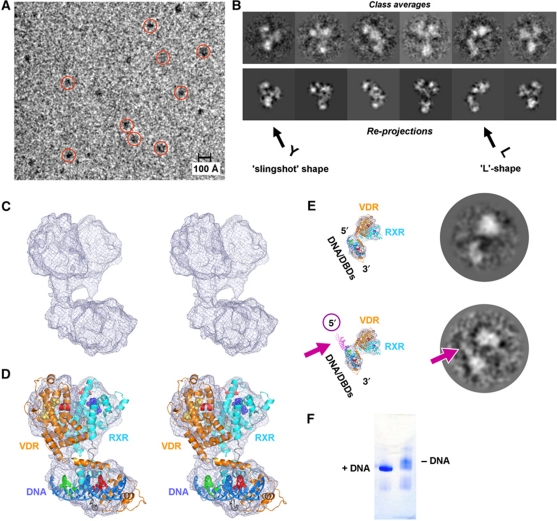
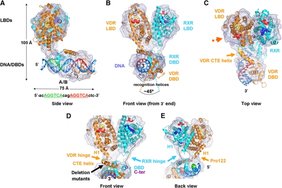
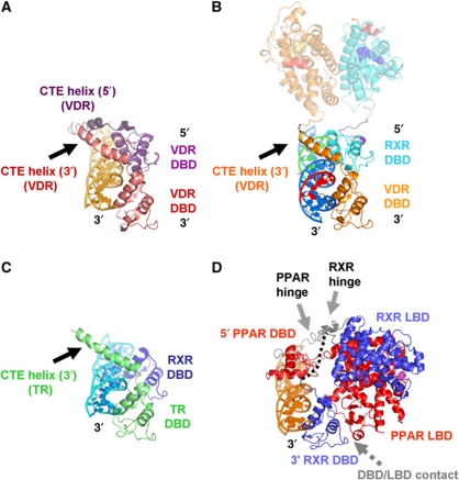
Transcription regulation by steroid hormones and other metabolites is mediated by nuclear receptors (NRs) such as the vitamin D and retinoid X receptors (VDR and RXR). Here, we present the cryo electron microscopy (cryo-EM) structure of the heterodimeric complex of the liganded human RXR and VDR bound to a consensus DNA response element forming a direct repeat (DR3). The cryo-EM map of the 100-kDa complex allows positioning the individual crystal structures of ligand- and DNA-binding domains (LBDs and DBDs). The LBDs are arranged perpendicular to the DNA and are located asymmetrically at the DNA 5'-end of the response element. The structure reveals that the VDR N-terminal A/B domain is located close to the DNA. The hinges of both VDR and RXR are fully visible and hold the complex in an open conformation in which co-regulators can bind. The asymmetric topology of the complex provides the structural basis for RXR being an adaptive partner within NR heterodimers, while the specific helical structure of VDR's hinge connects the 3'-bound DBD with the 5'-bound LBD and thereby serves as a conserved linker of defined length sensitive to mutational deletion.


Conflict of interest statement

The authors declare that they have no conflict of interest.

Figures

Figure 1
Figure 1
Structure determination and overall description of the RXR/VDR/DNA complex. (A) CCD image of a flash-frozen hydrated sample of the RXR/VDR/DNA complex; scale bar is 100 Å; recorded on a transmission electron microscope (see Materials and methods) under cryo conditions at a defocus of ∼5 μm at 200 kV (for processing, data were collected at a defocus of −2.0 up to −4.0 μm). Some particles are marked with red circles. A similarly good contrast is obtained also closer to focus (−2.5 μm defocus) when images are recorded at 100 kV (see Supplementary Figures S8 and S9). (B) Comparison of class averages (first lane, obtained by multivariate statistical analysis and classification) with the corresponding re-projections of the 3D reconstruction (second lane). The characteristic ‘L’ and ‘slingshot’ shapes can be recognized in the class averages (compare these views with the side and front views in Figure 2A and B). A comparison with raw particle images can be found in Supplementary Figure S10. (C) Stereo representation of the cryo-EM map revealing the global architecture of the RXR/VDR/DNA complex. (D) Stereo representation of the cryo-EM map with the fitted crystal structures of the individual RXR and VDR LBDs and DBDs, resulting in a molecular model of the full RXR/VDR/DNA complex. (E) Assignment of the DNA polarity within the complex via a 5′-extended DNA oligomer. Comparison of the class averages of the RXR/VDR complex with the 20-bp DNA (top lane) and with the longer DNA extended by 15 nucleotides on the 5′-end (bottom lane); the corresponding views with the modelled DNA are displayed on the left. The pink arrow indicates the additional density in the class average, which is observed at the expected 5′ position. These data confirm the localization of the DNA within the complex and the annotation of the DNA 5′–3′ polarity in the complex. (F) Native gel analysis of RXR/VDR in the presence (left) and absence (right) of the DNA. The sharp band in the presence of DNA indicates a conformational stabilization of the complex.
Figure 2
Figure 2
Structure of the RXR/VDR/DR3 DNA nuclear receptor complex. (A) Side view of the structure, with the 5′ DNA end on the left. The cryo-EM map is shown in magenta, the fitted DNA/DBD and LBD heterodimer parts are shown with their backbone secondary structure (models are derived from available crystal structures, see main text). The DNA is shown in blue with the first half-site of the response element in green and the second half-site in red. The DBDs of RXR (cyan) and VDR (orange) are bound on the back, and a density close to the recognition helix of VDR is attributable to the A/B domain of VDR (17 residues). The LBDs are oriented perpendicular to the DNA, anchored through the 5′ side of the response element. The dimensions of the complex are indicated as well as the sequence of the response element. (B) Front view of the complex as seen from the 3′-end of the DNA (rotated 90° with respect to the view in A). The VDR LBD is on the left whereas the RXR LBD is on the right. The DBDs sit side-on to the DNA and are rotated by ∼45° with respect to each other because of the three nucleotide spacer (DR3) between the half-sites. The ligands 1α,25-dihydroxyvitamin D3 and 9-cis retinoic acid for VDR and RXR are shown in yellow and blue van der Waals spheres. The area comprising helices H2, H3n and the β-sheet of VDR is marked with a star. Since the RXR partner is specific for co-activator binding, the site on VDR that includes trans-activation helix H12 (in red) is available for the recruitment of chromatin-modifying co-regulator proteins on the side oriented opposite to the DBDs. (C) Top view of the complex as seen along the pseudo two-fold axis through the interface of the LBDs (rotated 70° downwards with respect to the view in B). The C-terminal extension helix of VDR protruding from the DBD and crossing the DNA minor groove is indicated, and the LBD interface contact comprising helix 4, loop 8/9 of VDR and helix H7 of RXR are marked with a star. (D, E) Description of the hinge regions between the LBDs and DBDs of RXR (cyan) and VDR (orange). The map resolves the individual hinges of the complex, revealing that these are well defined, thus providing the connectivity between the LBD and DBD core domains. The hinges adopt a parallel arrangement and thus neither cross nor interact with each other. RXR has a 25 residue linker (dotted line) between the C-terminal end (dark red) of the DBD to the N-terminal end of helix H1 (dark red). For VDR, there is a direct connection between the C-terminal end (yellow) of the CTE and the N-terminal end of helix H1. The conserved Pro 122 marks the kink between the VDR CTE helix and helix H1. The representations in (D) and (E) are seen from the front (rotated 10° around the horizontal axis with respect to the view in B to better see the CTE helix) and from the back (rotated 150° around the vertical axis compared with D).
Figure 3
Figure 3
Comparison of the topology of NR complexes on the DNA. (A, B) Comparison of the crystal structure of the VDR homodimer DBD on a DR3 response element (PDB ID 1KB4; Shaffer and Gewirth, 2002, A) super-positioned to the DBD part of the cryo-EM structure of the RXR/VDR/DR3 complex (B, viewing angle and colour code as in Figure 2D; the LBD part is shown in faded colours for simplicity). The 5′ VDR DBD (violet) corresponds precisely to the RXR DBD in the RXR/VDR/DR3 complex, with the exception of the CTE helix (dark violet) that is absent in RXR (compare also with Figure 2D). This similarity provides an independent validation of the model of the RXR/VDR/DNA subcomplex and suggests that in homodimers the 5′ VDR plays the role of the 5′ RXR. (C) The DNA-bound TR DBD shows the same organization as the VDR DBD (viewing angle as in A and B), notably the CTE helix that protrudes from the DBD in the same direction coming from the 3′-bound DBD, suggesting that an RXR/TR/DR4 complex has a similar overall topology to RXR/VDR/DR3. (D) Comparison with the crystal structure of the PPAR/RXR/DR1 complex (PDB ID 3DZU; Chandra et al, 2008) super-positioned through the DNA and the 3′ DBDs of the cryo-EM structure of the RXR/VDR/DR3 complex (viewing angle and colour code as in AC; PPAR in blue, RXR in red, co-activator peptide in magenta; see also Supplementary Figure S6). The different response elements (DR3 versus DR1) lead to a reversal of the DNA polarity, but also to a transition from a side-on complex to an interaction from either sides of the DNA. The global architecture of the two complexes is also rather different with respect to the position of the LBDs which are perpendicular in the RXR/VDR complex while being in a closed conformation in the PPAR/RXR complex, resulting in an RXR DBD–PPAR LBD interaction (indicated in grey). In contrast, the RXR/VDR complex harbours only classical heterodimeric DBD–DBD and LBD–LBD interfaces. While the hinges are well defined in the cryo-EM structure of RXR/VDR (Figure 2), they are either not visible (black dotted line for the RXR hinge) or partially disordered (temperature factors of >90 Å2 in the PPAR hinge, the backbone is represented in grey).

Comment in

References

    1. Alimirah F, Vaishnav A, McCormick M, Echchgadda I, Chatterjee B, Mehta RG, Peng X (2010) Functionality of unliganded VDR in breast cancer cells: repressive action on CYP24 basal transcription. Mol Cell Biochem 342: 143–150 - PMC - PubMed
    1. Antony P, Sigüeiro R, Huet T, Sato Y, Ramalanjaona N, Rodrigues LC, Mouriño A, Moras D, Rochel N. (2010) Structure-function relationships and crystal structures of the vitamin D receptor bound 2 alpha-methyl-(20S,23S)- and 2 alpha-methyl-(20S,23R)-epoxymethano-1 alpha,25-dihydroxyvitamin D3. J Med Chem 53: 1159–1171 - PubMed
    1. Banerjee P, Chatterjee M (2003) Antiproliferative role of vitamin D and its analogs - a brief overview. Mol Cell Biochem 253: 247–254 - PubMed
    1. Bouillon R, Eelen G, Verlinden L, Mathieu C, Carmeliet G, Verstuyf A. (2006) Vitamin D and cancer. J Steroid Biochem Mol Biol 102: 156–162 - PubMed
    1. Bourguet W, Vivat V, Wurtz JM, Chambon P, Gronemeyer H, Moras D (2000) Crystal structure of a heterodimeric complex of RAR and RXR ligand-binding domains. Mol Cell 5: 289–298 - PubMed

Publication types

MeSH terms