. 2012 Oct 25;31(43):4599-608.
doi: 10.1038/onc.2011.587. Epub 2011 Dec 19.

Deubiquitination of EGFR by Cezanne-1 contributes to cancer progression

Affiliations

Deubiquitination of EGFR by Cezanne-1 contributes to cancer progression

F Pareja et al. Oncogene. .

Abstract

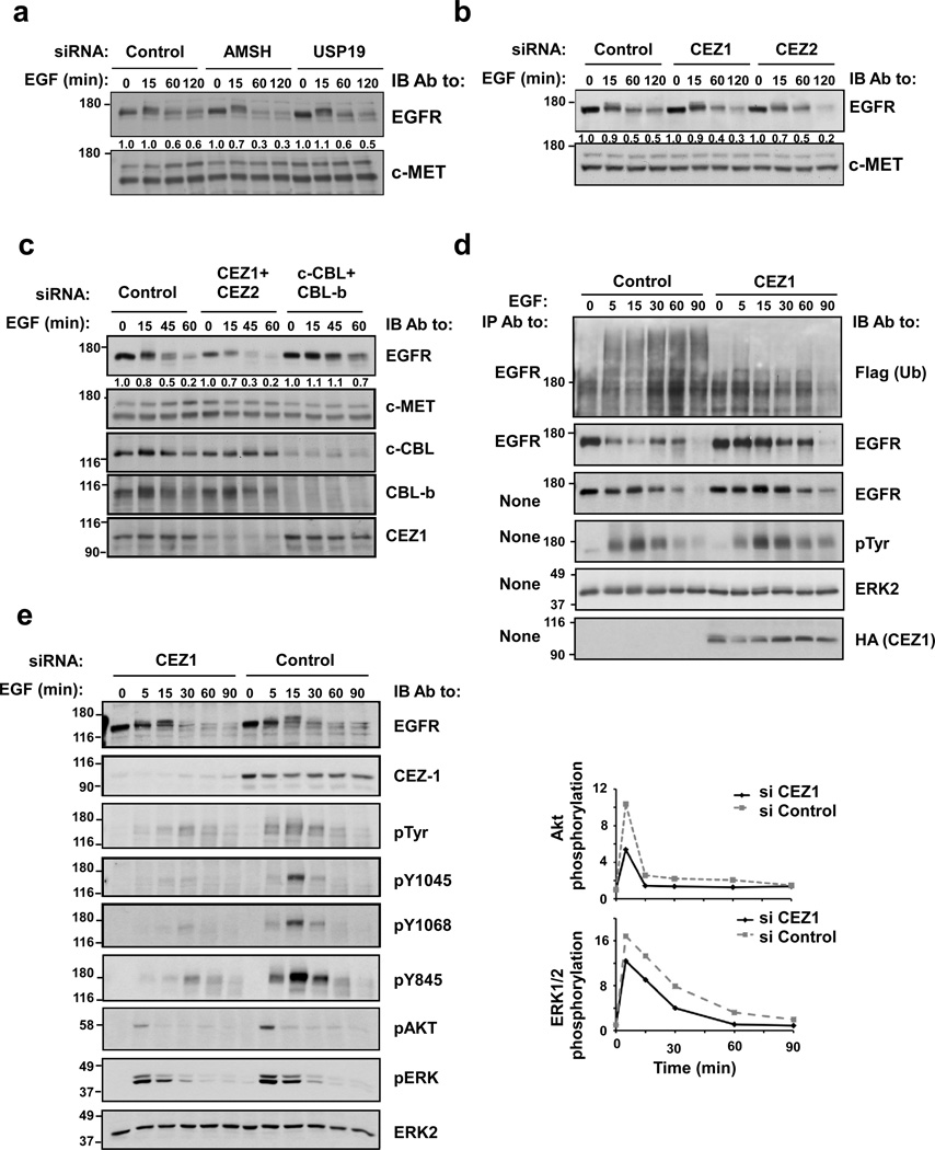
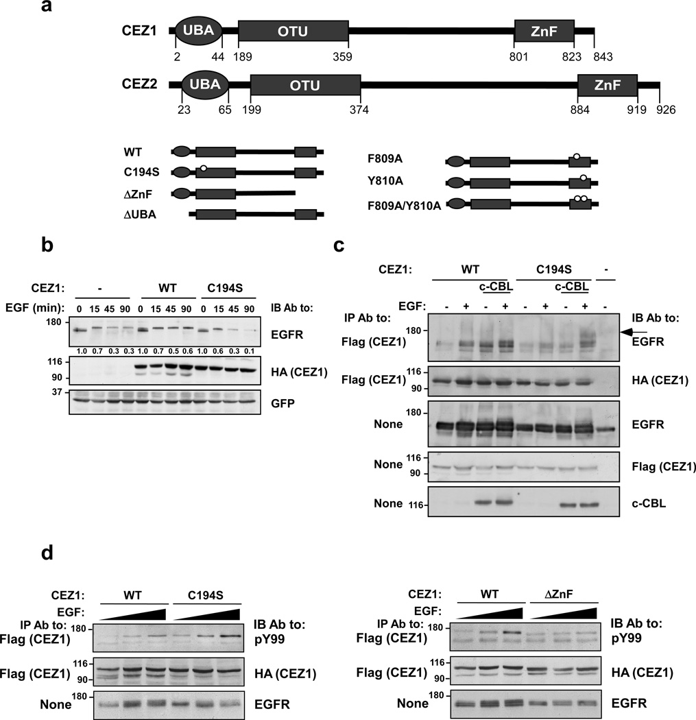
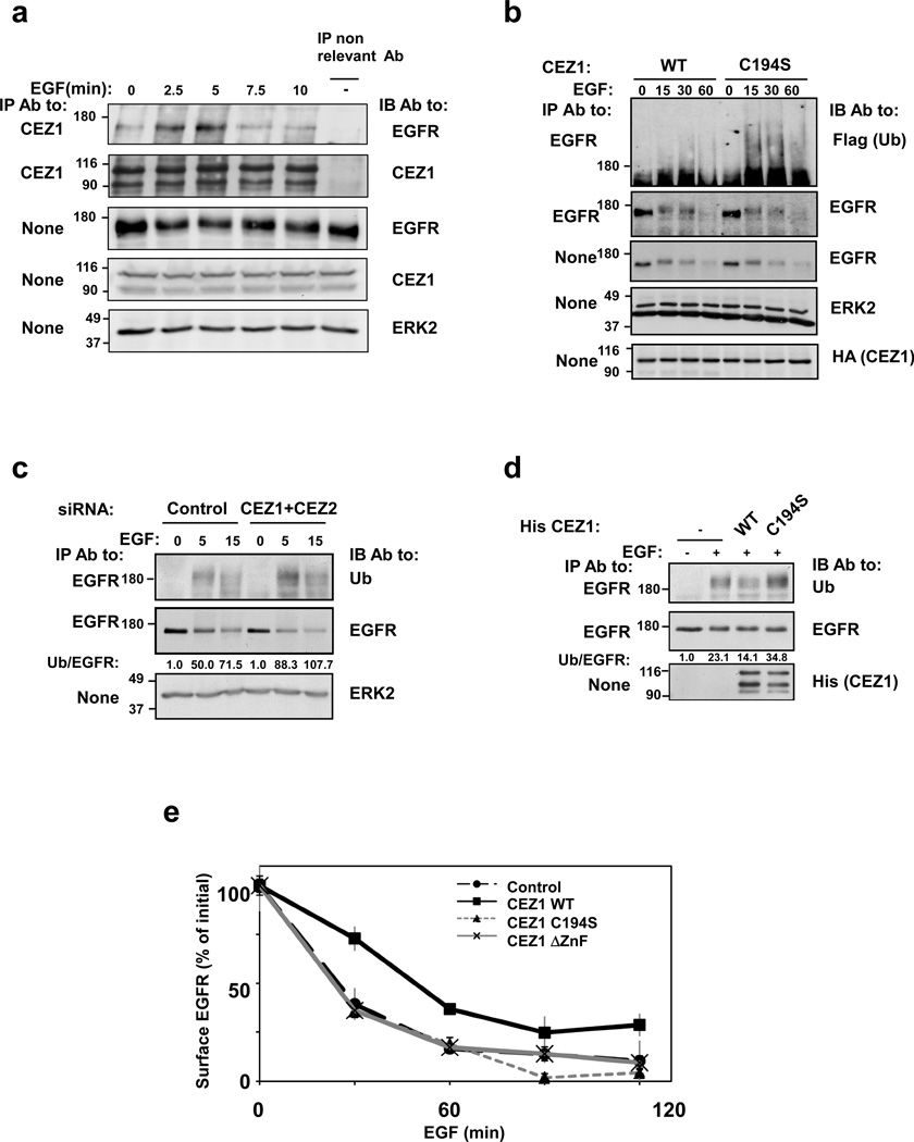
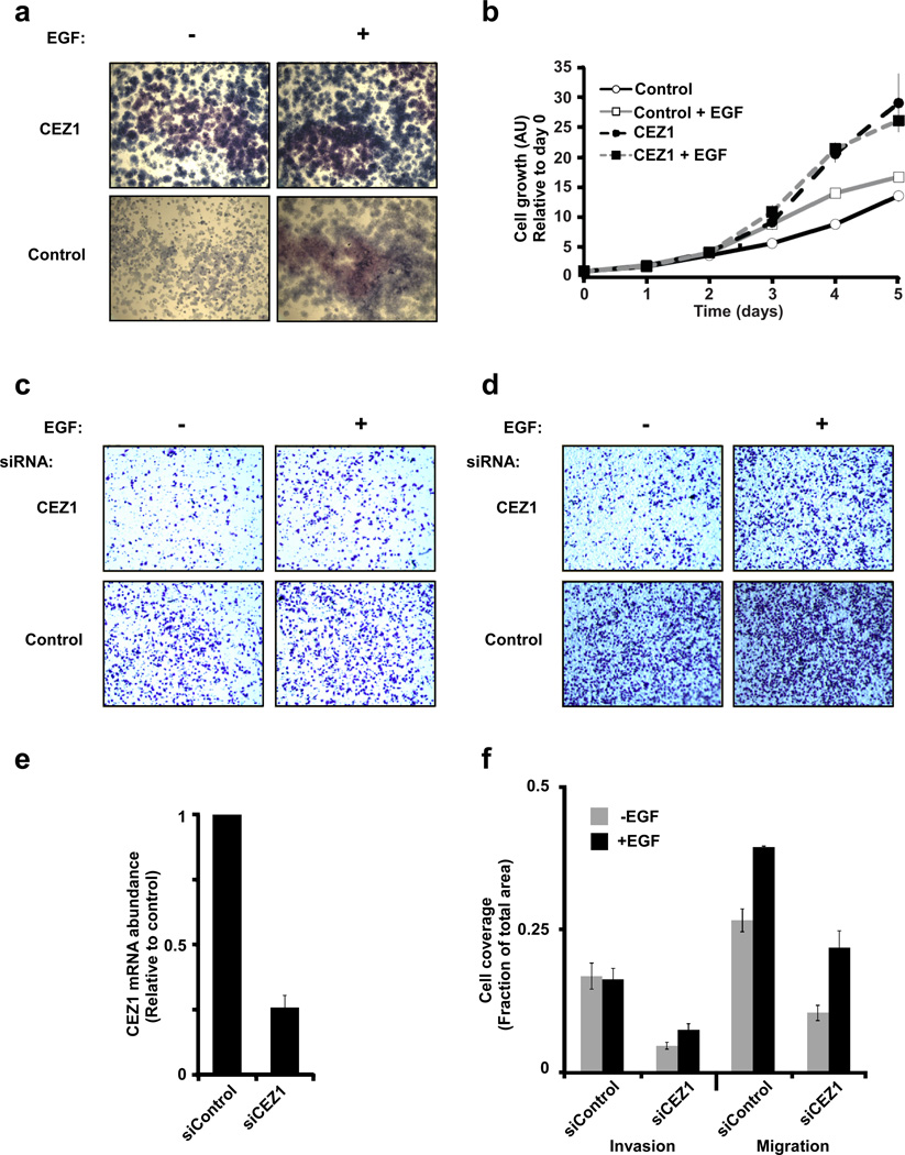
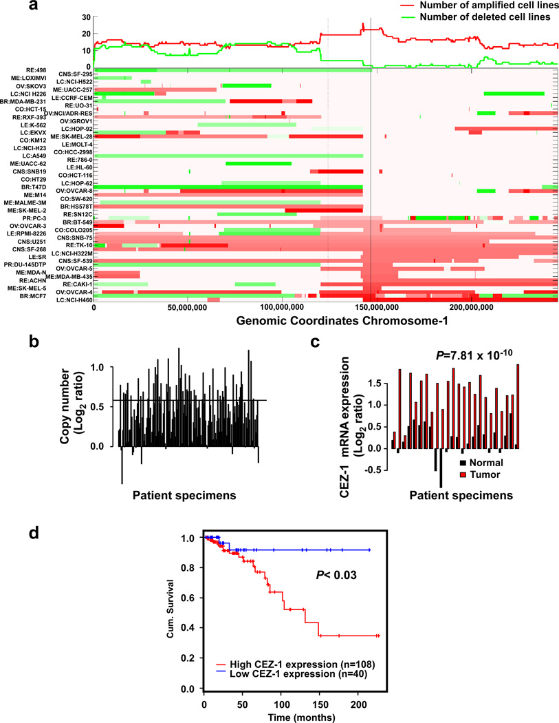
Once stimulated, the epidermal growth factor receptor (EGFR) undergoes self-phosphorylation, which, on the one hand, instigates signaling cascades, and on the other hand, recruits CBL ubiquitin ligases, which mark EGFRs for degradation. Using RNA interference screens, we identified a deubiquitinating enzyme, Cezanne-1, that opposes receptor degradation and enhances EGFR signaling. These functions require the catalytic- and ubiquitin-binding domains of Cezanne-1, and they involve physical interactions and transphosphorylation of Cezanne-1 by EGFR. In line with the ability of Cezanne-1 to augment EGF-induced growth and migration signals, the enzyme is overexpressed in breast cancer. Congruently, the corresponding gene is amplified in approximately one third of mammary tumors, and high transcript levels predict an aggressive disease course. In conclusion, deubiquitination by Cezanne-1 curtails degradation of growth factor receptors, thereby promotes oncogenic growth signals.


Conflict of interest statement

CONFLICT OF INTEREST

The authors declare no conflict of interest.

Figures

Figure 1
Figure 1. siRNA screens of human deubiquitinating enzymes identify Cezanne-1 and Cezanne-2 as EGFR-specific DUBs that enhance EGF signaling
(a) KB cells were transfected with siRNAs against AMSH and USP19. Forty-eight hours later, cells were stimulated with EGF (20 ng/ml), as indicated. Thereafter, lysates were immunoblotted (IB) with antibodies against EGFR and c-MET. The EGFR signals were quantified, normalized and indicated. (b) KB cells were transfected with siRNAs targeting Cezanne-1 and Cezanne-2, and cells were treated and analyzed as in a. (c) HeLa cells were transfected with combinations of siRNAs against Cezanne-1/Cezanne-2 and c-CBL/CBL-b. Forty-eight hours following transfection, cells were treated with EGF and whole cell lysates analyzed. (d) HeLa cells were transfected with a plasmid encoding Cezanne-1, or an empty plasmid, alongside with a plasmid encoding a Flag-tagged ubiquitin. Forty-eight hours later, cells were incubated with EGF (20 ng/ml) for the indicated intervals. Cell lysates and EGFR immunoprecipitates were analyzed as indicated. (e) HeLa cells were transfected with siRNAs targeting Cezanne-1, or with control oligonucleotides. Forty-eight hours later, cells were stimulated with EGF (20 ng/ml) for the depicted intervals. Thereafter, whole cell extracts were immunoblotted with the indicated antibodies, including antibodies to the phosphorylated forms of AKT and ERK, as well as antibodies specific to specific phosphorylated tyrosines of EGFR. Signal intensities of phosphorylated AKT and ERK2 were quantified, normalized and graphically presented (right panels). All panels presented in this figure were repeated at least thrice.
Figure 2
Figure 2. Cezanne-1 physically binds with and stabilizes EGFR and undergoes EGF-induced phosphorylation
(a) Schematic representations of the domain structures of Cezanne proteins. The following domains are depicted: Ubiquitin associated domain (UBA), otubain (OTU) and the Zinc Finger (ZnF). The mutants of Cezanne-1 used in this study (lower panel) include a point mutant in the OTU domain, a deletion mutant lacking the ZnF, a deletion of the UBA domain, and single and double point mutants within the ZnF. (b) HEK-293T cells were transfected with EGFR- and GFP-encoding plasmids, along with plasmids encoding Cezanne-1, either WT or C194S. After forty-eight hours, cells were incubated at 37°C with EGF (20 ng/ml). Cell lysates were immunoblotted with the indicated antibodies. This experiment was repeated twice. (c) HEK-293T cells were transfected with plasmids encoding EGFR, c-CBL or an empty plasmid. In addition, cells were transfected with the WT Cezanne-1 or C194S (Flag and HA tagged). Forty-eight hours later, cells were either stimulated with EGF (100 ng/ml) for 1 hour at 4°C, or left untreated. Thereafter, cleared cell extracts were subjected to immunoprecipitation (IP) with the indicated antibodies, or directly electrophoresed. An arrow indicates a form of EGFR whose mobility is retarded. This experiment was repeated thrice. (d) The indicated forms of HA-Flag-tagged Cezanne-1 were co-expressed together with EGFR in HEK-293T cells. Forty-eight hours after transfection, cells were treated for 10 minutes with increasing concentrations of EGF (0, 10 and 50 ng/ml) and cell lysates subjected to immunoprecipitation (IP) and immunoblotting (IB) using the indicated antibodies.
Figure 3
Figure 3. The catalytic and ubiquitin-binding functions of Cezanne-1 are essential for deubiquitination of EGFR and for inhibition of ligand-induced receptor downregulation
(a) MDA-MB-468 cells were stimulated with EGF (20 ng/ml) for the indicated time points. Whole cell lysates were subjected to immunoprecipitation with anti-Cezanne-1 antibodies, or with a non-relevant antibody. Immunoprecipitates were immunoblotted with antibodies against EGFR and Cezanne-1. Whole cell extracts were probed with antibodies to EGFR, Cezanne-1, and ERK2. (b) HeLa cells were transfected with plasmids encoding Cezanne-1, either WT or C194S, together with a Flag-tagged ubiquitin. Thereafter, cells were stimulated with EGF (20 ng/ml). Cell extracts were subjected to immunoprecipitation (IP) and/or immunoblotting (IB) with the indicated antibodies. (c) HeLa cells were transfected with a mixture of siRNAs against Cezanne-1 and Cezanne-2, or with control oligonucleotides. Forty-eight hours after transfection, cells were incubated with EGF (20 ng/ml) and lysates were subjected to immunoprecipitation and immunoblotting as indicated. Signals were quantified, normalized and the ubiquitin/EGFR ratio indicated. (d) HeLa cells were stimulated for 15 minutes with EGF (20 ng/ml). Subsequently, cells were lysed and their extracts subjected to immunoprecipitation with anti-EGFR antibodies. Thereafter, EGFR immunoprecipitates were incubated for 1 hour at 37°C with the WT or C194S forms of a His-tagged Cezanne-1, previously purified from bacteria. The immunoprecipitates were subsequently immunoblotted with antibodies against ubiquitin and EGFR; signals were quantified, normalized and their ratio presented. (e) HeLa cells were transfected with plasmids encoding the indicated forms of Cezanne-1, or with an empty vector. Thereafter, cells were stimulated with EGF (2 ng/ml) for the indicated time intervals. Next, EGFR downregulation was assayed using a radioactive EGF. Averages of triplicates and S.D. values (bars) are presented. All experiments presented in this figure were repeated at least thrice.
Figure 4
Figure 4. The Zinc Finger of Cezanne-1 binds with the Asp58-centered surface of ubiquitin
(a) HEK-293T cells were transfected with plasmids encoding Flag-tagged Cezanne-1 (WT and FY/AA). Forty-eight hours later, lysates were incubated for 1 hour at 4°C with immobilized GST-ubiquitin, either WT, I44A or D58A. Pull-downs were stained or probed with an antibody to Flag. The Input lane represents 10% of the amount of cell extract used per lane. This experiment was repeated twice. (b) Lysates of HEK-293T cells transiently expressing Cezanne-1 were incubated for 1 hour at 4°C with an immobilized GST-I44A-Ub. Pull-downs were stained or probed with an antibody to Flag. The input extracts were electrophoresed for reference. This experiment was repeated twice. (c) HEK-293T cells were transfected with plasmids encoding EGFR and the indicated forms of Cezanne-1. Forty-eight hours later, cells were stimulated with EGF (20 ng/ml). Cleared extracts were probed as indicated. (d) HeLa cells were transfected with the indicated forms of Cezanne-1. After 48 hours cells were stimulated with EGF (20 ng/ml). Whole extracts were probed as indicated.
Figure 5
Figure 5. Cezanne-1 associates with cell proliferation and migration
(a and b) HeLa cells stably overexpressing Cezanne-1 (or an empty plasmid) were plated, allowed to adhere overnight, and stimulated with EGF (20 ng/ml). Subsequently, the cells were allowed to grow for 7 days, fixed and stained with Giemsa prior to image capturing (a). Alternatively, cells were stimulated with EGF (20 ng/ml) and grown for the indicated intervals prior to an assay that determined cell growth (b). The results represent average±S.D. values of quadruplets. (c and d) MDA-MB-231 cells were transfected with the indicated siRNA oligonucleotides and 24 hours later they were seeded in the upper compartment of an Invasion (c) or a Transwell (d) chamber. The lower compartment of each chamber was filled with either serum-containing medium or the same medium supplemented with EGF (10 ng/ml). Eighteen hours later, cells that migrated to the lower side of the filter were fixed, permeabilized and stained. (e) MDA-MB-231 cells were transfected with siRNA oligonucleotides as in c and d, and expression levels of Cezanne-1 were determined using a preparation of mRNA and PCR. (f) Quantification of cell invasion and migration (panels c and d) by measuring the fraction of the lower face of the intervening filter which was covered by migrating cells. Bars show the means and S.D. values of three determinations.
Figure 6
Figure 6. Cezanne-1 associates with advanced malignancy of breast lesions
(a) Photomicrographs presenting scores of Cezanne-1 expression in breast cancer specimens, qualified as strong, moderate, weak and negative. (b) The abundance of Cezanne-1 is compared to the following disease subgroups (the respective numbers of cases are indicated): normal breast, benign disease, ductal and lobular carcinoma in situ (DCIS/LCIS), invasive ductal carcinoma (IDC), invasive lobular carcinoma (ILC), and other invasive carcinomas. Statistical p values were calculated (Chi square test): * represents p <0.01, and ** represents p <0.001.
Figure 7
Figure 7. Genomic amplification and high mRNA levels of Cezanne-1 in mammary tumors, and their association with shorter patient survival time
(a) Heatmap representation of chromosome 1 aberrations, based on the NCI60 set of cancer cell lines. The x-axis represents genomic coordinates, including the centromer (dashed vertical line) and CEZ1 (solid line). The top panel summarizes the number of amplified (or deleted) cell lines. Each row in the bottom panel represents a cell line. The left axis lists all lines, sorted according to CEZ1 copy number. Amplifications are marked in red, deletions in green. The scale of colors is based on the log ratio between the respective cell line and a normal sample (log ratio of zero is shown in white). A CNV (at 149,400,000) deleted in 26 samples was not included. (b) DNA copy number of CEZ1 in tumors from breast cancer patients. The x-axis represents 173 patients, 56 of them have gained at least 1 extra copy of CEZ1. The y-axis represents log2 ratio of SNP intensity using paired blood-derived normal cells as control. The horizontal line represents log2 ratio value for 3 copies. (c) mRNA abundance of Cezanne-1 in pairs of a breast tumor and adjacent normal tissues from 24 breast cancer patients. The statistical p value, determined by using the paired two-sample T test, is indicated. (d) Kaplan-Meyer survival analysis was performed on 148 breast cancer patients. All patients were divided into high and low CEZ1 expression groups. The cutoff was optimized to achieve low p-value.

References

    1. Avraham R, Yarden Y. Feedback regulation of EGFR signalling: decision making by early and delayed loops. Nat Rev Mol Cell Biol. 2010;12:104–117. - PubMed
    1. Bowers K, Piper SC, Edeling MA, Gray SR, Owen DJ, Lehner PJ, et al. Degradation of endocytosed epidermal growth factor and virally ubiquitinated major histocompatibility complex class I is independent of mammalian ESCRTII. J Biol Chem. 2006;281:5094–5105. - PubMed
    1. Caswell PT, Chan M, Lindsay AJ, McCaffrey MW, Boettiger D, Norman JC. Rab-coupling protein coordinates recycling of alpha5beta1 integrin and EGFR1 to promote cell migration in 3D microenvironments. J Cell Biol. 2008;183:143–155. - PMC - PubMed
    1. Cheng KW, Lahad JP, Kuo WL, Lapuk A, Yamada K, Auersperg N, et al. The RAB25 small GTPase determines aggressiveness of ovarian and breast cancers. Nat Med. 2004;10:1251–1256. - PubMed
    1. Courbard JR, Fiore F, Adelaide J, Borg JP, Birnbaum D, Ollendorff V. Interaction between two ubiquitin-protein isopeptide ligases of different classes, CBLC and AIP4/ITCH. J Biol Chem. 2002;277:45267–45275. - PubMed

Publication types