. 2012 Sep 27;31(39):4317-32.
doi: 10.1038/onc.2011.593. Epub 2011 Dec 19.

Inhibition of cell migration by PITENINs: the role of ARF6

Affiliations

Inhibition of cell migration by PITENINs: the role of ARF6

B Miao et al. Oncogene. .

Abstract

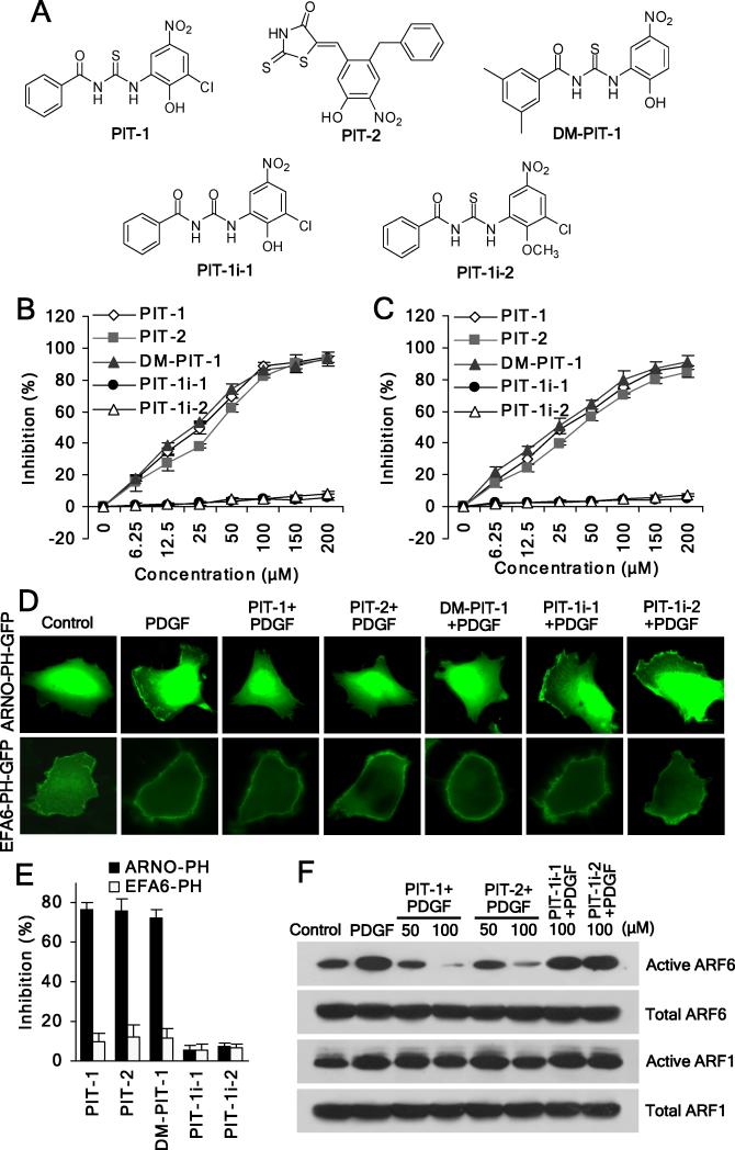
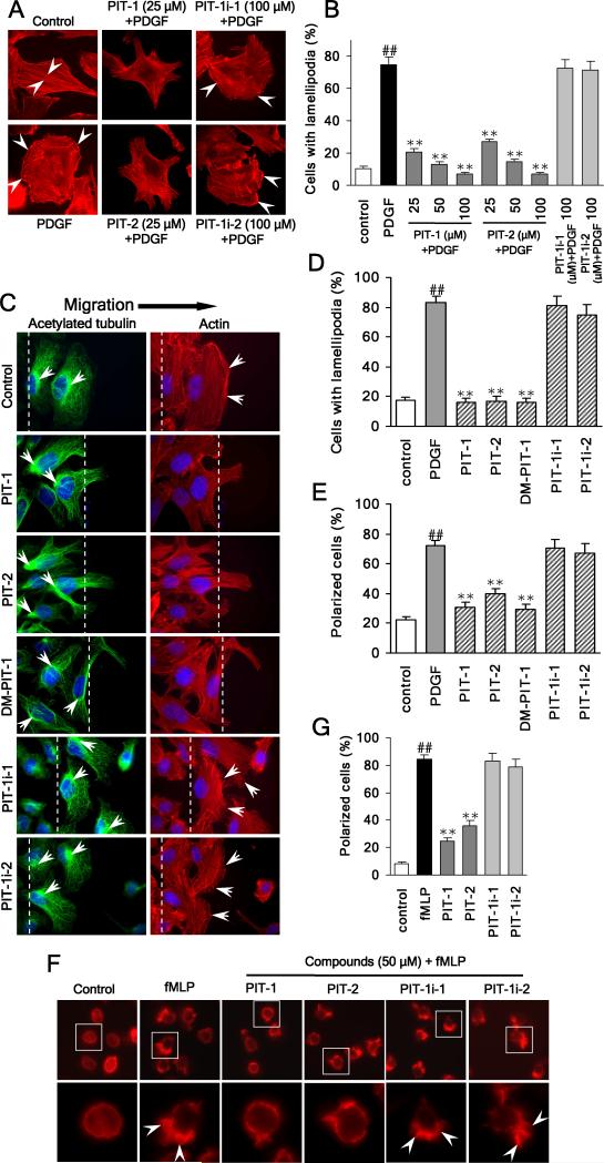
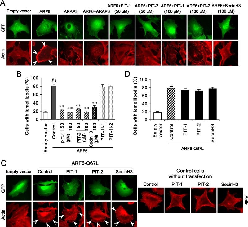
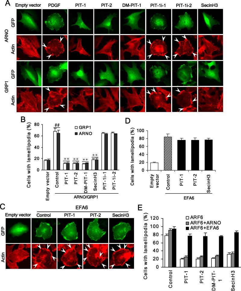
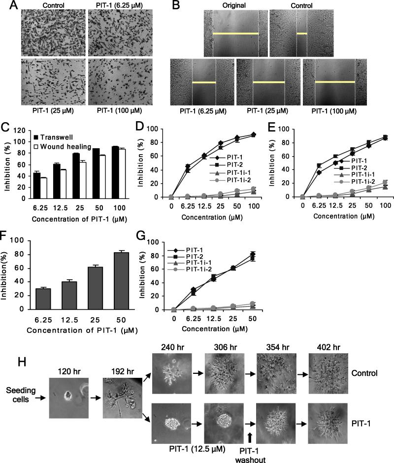
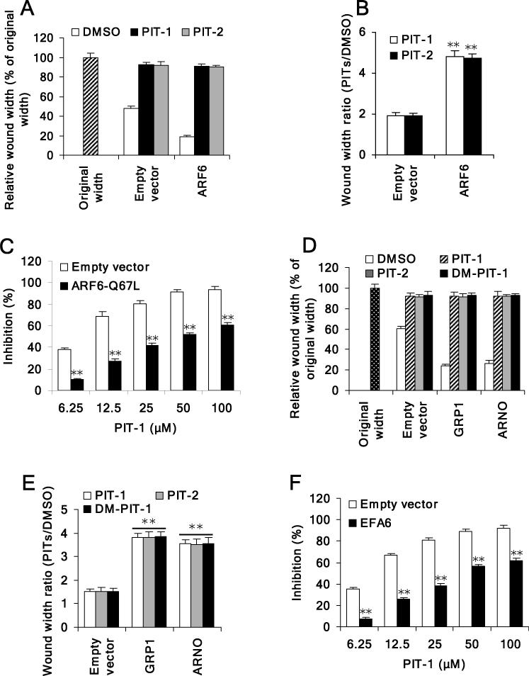
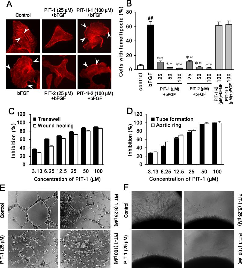
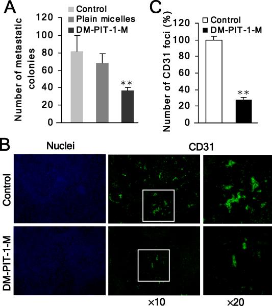
We have reported previously the development of small-molecule phosphatidylinositol-3,4,5-trisphosphate (PIP3) antagonists (PITs) that block pleckstrin homology (PH) domain interaction, including activation of Akt, and show anti-tumor potential. Here we show that the same molecules inhibit growth factor-induced actin remodeling, lamellipodia formation and, ultimately, cell migration and invasion, consistent with an important role of PIP3 in these processes. In vivo, a PIT-1 analog displays significant inhibition on tumor angiogenesis and metastasis. ADP ribosylation factor 6 (ARF6) was recently identified as an important mediator of cytoskeleton and cell motility, which is regulated by PIP3-dependent membrane translocation of the guanine nucleotide exchange factors (GEFs), such as ADP-ribosylation factor nucleotide binding site opener (ARNO) and general receptor for 3-phosphoinositides (GRP1). We demonstrate that PITs inhibit PIP3/ARNO or GRP1 PH domain binding and membrane localization, resulting in the inhibition of ARF6 activation. Importantly, we show that expression of the constitutively active mutant of ARF6 attenuates inhibition of lamellipodia formation and cell migration by PITs, confirming that inhibition of ARF6 contributes to inhibition of these processes by PITs. Overall, our studies demonstrate the feasibility of developing specific small-molecule targeting PIP3 binding by PH domains as potential anticancer agents that can simultaneously interfere with cancer development at multiple points.


Figures

Figure 1
Figure 1. PIT-1 inhibits PIP3 binding to GRP1/ARNO PH domains, and suppresses ARF6 activation
(A) Structures of the PITs and inactive analogs. (B,C) PITs inhibit PIP3/GRP1 or ARNO PH domain binding. TMR-conjugated PIP3 (60 nM) was incubated with GRP1 (B) or ARNO (C) PH domain (100 nM) in the presence of PITs for 40 min, followed by FP measurement. (D,E) PITs inhibit plasma membrane translocation of ARNO, but not EFA6 PH domain. After transfection, cells were serum starved and incubated with 50 μM PITs or inactive analogues for 2 hr, followed by stimulation with 100 ng/ml PDGF for 5 min. The translocation was analyzed using a fluorescent microscope. The representative fluorescent images are shown (D). The number of cells with membrane located GFP was quantitated by counting in five random fields and the inhibition was calculated (E). (F) PITs significantly inhibit ARF6 activation. SUM159 cells were serum starved and treated with compounds for 2 hr, followed by stimulation with 100 ng/ml PDGF for 15 min. ARF6 and ARF1 activation assays were performed as described in the Methods section.
Figure 2
Figure 2. PIT-1 inhibits lamellipodia formation and cell polarization
(A) PITs inhibit lamellipodia formation in cancer cells. SUM159 cells were serum starved and treated with compounds for 2 hr, followed by PDGF (100 ng/ml) stimulation for 15 min. Then cells were stained and analyzed using fluorescent microscopy. The representative fluorescent images are shown (A, the stress fibers and lamellipodia are indicated by arrowheads). The number of cells with significant lamellipodia was counted randomly in five fields and the percentage was calculated (B). (C-E) PITs inhibit cell polarization and lamellipodia formation. Wounds were generated in SUM159 cell monolayers. Then SUM159 cells were serum starved and treated with compounds for 2 hr, followed by stimulation with 100 ng/ml PDGF for 15 min. Then cells were fixed and stained with TRITC-phalloidin for F-actin (red), acetylated tubulin antibody (green) for microtubule organization center (MTOC), and Hoechst for nuclei (blue). Cell polarization was evaluated by analyzing the relative location of the MTOC and nuclei. The cell was considered polarized if MTOC was observed to the wound side of the nuclei. The representative fluorescent images are shown (C, The wound edge is indicated with white lines and the actin ruffles and the MTOC are marked with white arrowheads). The number of polarized cells (E), as well as cells with significant lamellipodia (D) was counted in five random fields under a fluorescent microscope and the percentage was calculated. (F,G) PITs inhibit fMLP stimulated HL-60 cell polarization. Differentiated HL-60 cells were treated with compounds at indicated concentrations for 2 hr, followed by stimulation with 100 nM fMLP for 3 min. Actin was stained and analyzed using fluorescent microscopy. The representative fluorescent images are shown (F, the leading edge of the polarized cells with lamellipodia is indicated by arrowheads). The number of polarized cells (there is obvious leading edge with lamellipodia) was counted randomly in five fields and the percentage was calculated (G). ##P<0.01 compared with control group, **P<0.01 compared with PDGF/fMLP group.
Figure 3
Figure 3. PIT-1 suppresses ARF6 but not ARF6-Q67L induced lamellipodia formation
(A,B) PITs inhibit lamellipodia formation induced by ARF6 overexpression. SUM159 cells were transfected with ARF6 or ARAP3, or co-transfected with both. Then cells were serum starved and treated with compounds for 2 hr, followed by stimulation with 100 ng/ml PDGF for 15 min. Cells were stained and analyzed with fluorescent microscopy. The representative fluorescent images are shown (A, the lamellipodia are indicated by arrowheads). The number of cells with significant lamellipodia was counted randomly in five fields and the percentage was calculated (B) (C,D) PITs fail to inhibit lamellipodia formation induced by ARF6-Q67L overexpression. SUM159 cells were transfected with ARF6-Q67L. Then cells were treated with compounds for 2 hr, followed by staining and fluorescent microscopy analysis. The representative fluorescent images are shown (C) and the number of cells with significant lamellipodia was counted in five random fields and the percentage was calculated (D). ##P<0.01 compared with empty vector group, **P<0.01 compared with ARF6 overexpression control groups.
Figure 4
Figure 4. PIT-1 suppresses ARNO/GRP1 but not EFA6 induced lamellipodia formation
(A,B) PITs inhibit lamellipodia formation induced by ARNO/GRP1 overexpression. SUM159 cells were transfected with full length ARNO or GRP1. Then cells were serum starved and treated with compounds for 2 hr, followed by stimulation with 100 ng/ml PDGF for 15 min. Cells were stained and analyzed with fluorescent microscopy. The representative fluorescent images are shown (A). The number of cells with significant lamellipodia was counted in five random fields and the percentage was calculated (B). (C,D) PITs fail to inhibit lamellipodia formation induced by EFA6 overexpression. SUM159 cells were transfected with EFA6. Then cells were treated with compounds for 2 hr, followed by staining and fluorescent microscopy analysis. The representative fluorescent images are shown (C) and the number of cells with significant lamellipodia was counted in five random fields and the percentage was calculated (D). (E) EFA6 but not ARNO can overcome inhibition of ARF6 by PITs. SUM159 cells were transfected with ARF6 or co-transfected with ARF6 and EFA6 or ARNO. Then cells were treated with compounds for 2 hr, followed by staining and fluorescent microscopy analysis. The number of cells with significant lamellipodia was counted in five random fields and the percentage was calculated. ##P<0.01 compared with empty vector group, **P<0.01 compared with ARNO/GRP1 overexpression control groups.
Figure 5
Figure 5. PIT-1 inhibits cancer cell migration and invasion
(A) PIT-1 inhibits cancer cell migration in a transwell assay. SUM159 cells were treated with PIT-1 for 8 hr. The cells on the lower side of chamber were stained and the representative images are shown, then cells were lysed and colorimetric determination was made at 595 nm. (B) PIT-1 inhibits cancer cell migration in a wound healing assay. A scratch was introduced into a monolayer of SUM159 cells, followed by treatment with PIT-1 for 8 hr. The width of wounded cell monolayer was measured in five random fields, and the representative images are shown. (C) Quantitation of the data from the assays in (A) and (B). (D,E) PIT-1 and PIT-2, but not PIT-1i-1 and PIT-1i-2, inhibit SUM159 cell migration in transwell (D) and wound healing (E) assays. (F) PIT-1 inhibits cancer cell invasion. SUM159 cells were seeded on a matrigel pre-coated transwell membrane, and the treatment and analysis are similar with transwell assay described above. (G) PIT-1 and PIT-2, but not PIT-1i-1 and PIT-1i-2, inhibit cancer cell invasion through a matrigel-coated membrane. (H) PIT-1 reversibly inhibits acquisition of the invasive phenotype of SUM159 cells in a 3-D matrigel matrix. SUM159 cells were seeded in matrigel and incubated for 192 hr, followed by the treatment with PIT-1 (12.5 μM) for 114 hr. Subsequently, PIT-1 was washed out and cells were incubated for additional 96 hr. The representative images are shown.
Figure 6
Figure 6. PIT-1 suppresses ARF6 or GRP1/ARNO induced cell migration
(A,B) PITs inhibit ARF6-induced cell migration. SUM159 cells were transfected with ARF6. Scratch wound was generated in cell monolayer, followed by treatment with PITs (50 μM) for 8 hr. The width of wounded cell monolayers was measured in five random fields and expressed as % of original width (A). The wound width ratio (PITs treated groups/DMSO control group) was calculated (B). (C) Overexpression of ARF6-Q67L attenuates inhibition of cell migration led by PIT-1. SUM159 cells were transfected with ARF6-Q67L. Scratch wound was generated in cell monolayer, followed by treatment with compounds for 8 hr. The width of wounded cell monolayer was measured in five random fields, and the inhibition was calculated. (D,E) PITs inhibit GRP1/ARNO-induced cell migration. SUM159 cells were transfected with full length GRP1 or ARNO. Scratch wound was generated in cell monolayer, followed by treatment with PITs (50 μM) for 8 hr. The width of wounded cell monolayers was measured in five random fields and expressed as % of original width (D). The wound width ratio (PITs treated groups/DMSO control group) was calculated (E). (F) Overexpression of EFA6 attenuates inhibition of cell migration by PIT-1. SUM159 cells were transfected with EFA6. Scratch wound was generated in cell monolayer, followed by treatment with compounds for 8 hr. The width of wounded cell monolayer was measured in five random fields, and the inhibition was calculated. *P<0.05, **P<0.01 compared with empty vector group.
Figure 7
Figure 7. PIT-1 inhibits angiogenesis in vitro
(A,B) PITs inhibit lamellipodia formation in HUVEC cells. Cells were serum starved and treated with compounds for 2 hr, followed by bFGF (10 ng/ml) stimulation for 15 min. Then cells were stained and analyzed as described above (A, the stress fibers and lamellipodia are indicated by arrowheads). The number of cells with significant lamellipodia was counted in five random fields and the percentage was calculated (B). (C) PIT-1 inhibits endothelial cell (HUVEC) migration in transwell and wound healing assays. The cells were treated and analyzed as described above. (D-F) PIT-1 inhibits tube formation and microvessel outgrowth. (D) Quantitation of the effects of PIT-1 on endothelial cell tube formation and microvessel outgrowth from (E) and (F). (E) PIT-1 inhibits tube formation with endothelial cells. HUVEC cells were seeded on matrigel and treated with PIT-1 for 8 hr. The representative images are shown. The tube formation was assessed by counting the number of closed tubes in five random fields from each well and the inhibition was calculated. (F) PIT-1 suppresses microvessel outgrowth in the aortic ring sprouting experiment. The rat aortic rings were embedded in matrigel and treated with PIT-1 for 6 days, followed by counting the number of microvessel outgrowths under a microscope. The representative images are shown and the inhibition was calculated. ##P<0.01 compared with control group, **P<0.01 compared with bFGF group.
Figure 8
Figure 8. DM-PIT-1 inhibits in vivo cancer angiogenesis and metastasis
(A) Administration of DM-PIT-1-M (5 mg/kg/day) suppresses pulmonary metastasis formation with B16-F10 melanoma cells. After 5-day drug administration, the mean number of metastatic colonies was counted. (B,C) DM-PIT-1-M administration suppresses in vivo angiogenesis in tumors evaluated using IH assay with anti-CD31 antibody (green). Nuclei were stained with Hoechst (blue). The representative fluorescent images are shown (B) and the number of CD31 foci was counted in five random fields and expressed as % of control (C). **P<0.01 compared with control group.

References

    1. Avraamides CJ, Garmy-Susini B, Varner JA. Integrins in angiogenesis and lymphangiogenesis. Nat Rev Cancer. 2008;8:604–17. - PMC - PubMed
    1. Balana ME, Niedergang F, Subtil A, Alcover A, Chavrier P, Dautry-Varsat A. ARF6 GTPase controls bacterial invasion by actin remodelling. J Cell Sci. 2005;118:2201–10. - PubMed
    1. Cantley LC. The phosphoinositide 3-kinase pathway. Science. 2002;296:1655–7. - PubMed
    1. Caumont AS, Vitale N, Gensse M, Galas MC, Casanova JE, Bader MF. Identification of a plasma membrane-associated guanine nucleotide exchange factor for ARF6 in chromaffin cells. Possible role in the regulated exocytotic pathway. J Biol Chem. 2000;275:15637–44. - PubMed
    1. Cavenagh MM, Whitney JA, Carroll K, Zhang C, Boman AL, Rosenwald AG, et al. Intracellular distribution of Arf proteins in mammalian cells. Arf6 is uniquely localized to the plasma membrane. J Biol Chem. 1996;271:21767–74. - PubMed

Publication types

MeSH terms