. 2012 Apr;40(8):3623-40.
doi: 10.1093/nar/gkr1156. Epub 2011 Dec 17.

A conserved RpoS-dependent small RNA controls the synthesis of major porin OmpD

Affiliations

A conserved RpoS-dependent small RNA controls the synthesis of major porin OmpD

Kathrin S Fröhlich et al. Nucleic Acids Res. 2012 Apr.

Abstract

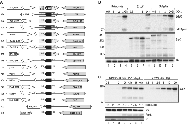
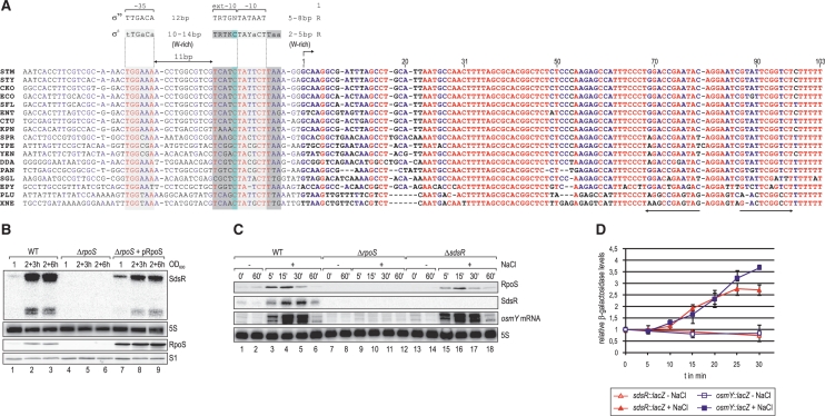
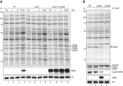
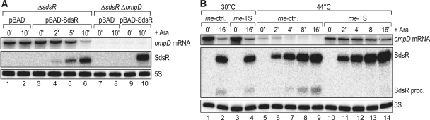
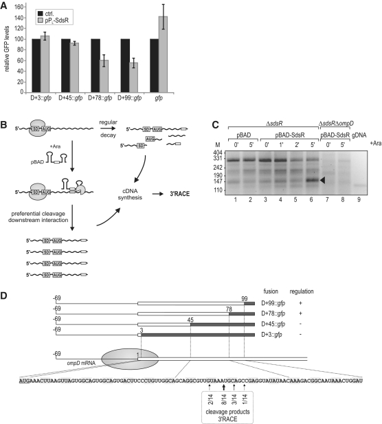
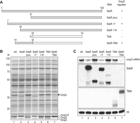
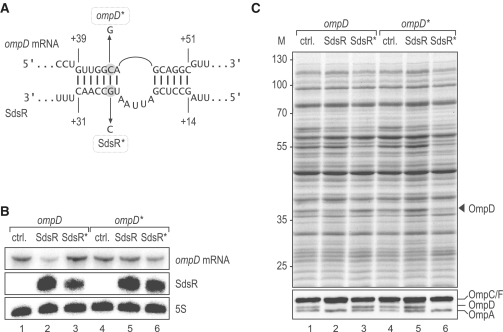
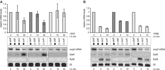
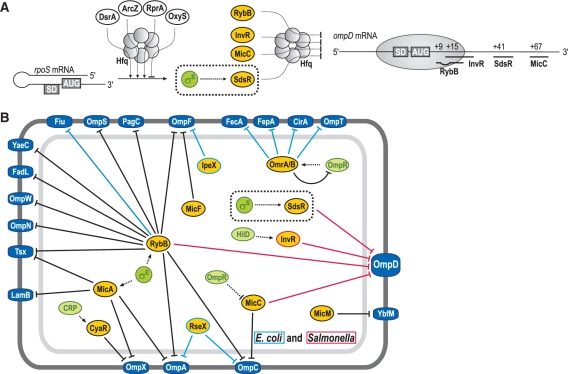
A remarkable feature of many small non-coding RNAs (sRNAs) of Escherichia coli and Salmonella is their accumulation in the stationary phase of bacterial growth. Several stress response regulators and sigma factors have been reported to direct the transcription of stationary phase-specific sRNAs, but a widely conserved sRNA gene that is controlled by the major stationary phase and stress sigma factor, σ(S) (RpoS), has remained elusive. We have studied in Salmonella the conserved SdsR sRNA, previously known as RyeB, one of the most abundant stationary phase-specific sRNAs in E. coli. Alignments of the sdsR promoter region and genetic analysis strongly suggest that this sRNA gene is selectively transcribed by σ(S). We show that SdsR down-regulates the synthesis of the major Salmonella porin OmpD by Hfq-dependent base pairing; SdsR thus represents the fourth sRNA to regulate this major outer membrane porin. Similar to the InvR, MicC and RybB sRNAs, SdsR recognizes the ompD mRNA in the coding sequence, suggesting that this mRNA may be primarily targeted downstream of the start codon. The SdsR-binding site in ompD was localized by 3'-RACE, an experimental approach that promises to be of use in predicting other sRNA-target interactions in bacteria.


Figures

Figure 1.
Figure 1.
(A) Genomic context of the sdsR gene in various Enterobacteria. Synteny analysis of the sdsR/sraC genes in various enterobacterial species revealed partial conservation of the locus. In all cases sdsR is positioned downstream of yebY or homologues; the flanking gene at the 3′-end is variable. Distances to flanking genes are indicated in bp. STM: Salmonella typhimurium; STY: Salmonella typhi; CKO: Citrobacter koseri; ECO: Escherichia coli; SFL: Shigella flexneri; ENT: Enterobacter; CTU: Cronobacter turicensis; KPN: Klebsiella pneumoniae; SPR: Serratia proteamaculans; YPE: Yersinia pestis; YEN: Yersinia enterolitica; DDA: Dickeya dadantii; PAN: Pantoea ananatis; SGL: Sodalis glossinidius; EPY: Erwinia pyrifoliae; PLU: Photorhabdus luminescens; XNE: Xenorhabdus nematophila. (B) Expression levels of SdsR and SraC sRNAs in Salmonella, E. coli and Shigella. Northern blot analysis of total RNA isolated from wild-type Salmonella (JVS-1574), E. coli (JVS-5105) and Shigella (JVS-0012) cells grown to OD600 of 0.5, 1.0, 2.0 and 3 h after cells had reached an OD600 of 2.0. SdsR and SraC sRNAs were detected by radio-labelled oligo probes directed against the conserved sRNA sequences. 5S rRNA levels were determined to confirm equal loading. (C) SdsR copy number over growth. SdsR levels at various timepoints (OD600 of 0.5, 1.0, 2.0 and 2, 3, 4 or 6 h after cells had reached an OD600 of 2.0) in wild-type Salmonella were compared by northern blotting to signals of in vitro transcribed SdsR in indicated amounts. SdsR was detected using a riboprobe; probing for 5S RNA confirmed equal loading. Expression of RpoS at different growth stages was determined by western blotting. Detection of ribosomal protein S1 was used as loading control.
Figure 2.
Figure 2.
(A) Non-redundant alignment of the sdsR gene including the upstream promoter region. All nucleotides are coloured regarding their degree of conservation (red: high conservation; blue: partial conservation; black: little or no conservation). σ70 or σS-specific promoter consensus motifs (61) are indicated above the alignment (W: A/T; R: A/G; Y: C/T; K: T/G); the putative −10 and the −35 sites of the sdsR promoter are boxed in light grey, σS-specific extensions of the −10 element are marked in dark grey and the conserved cytosine residue at position −13 is boxed in light blue. ‘+1’ marks the transcriptional start site, and the transcribed SdsR sequence is given in bold letters. The ρ-independent terminator is indicated by arrows. (B) SdsR sRNA is not detectable in the absence of RpoS. SdsR levels were determined by northern blotting of RNA isolated at indicated times over growth from Salmonella wild-type and rpoS mutant cells (JVS-5487) carrying either a control vector (pRH800) or a plasmid constitutively expressing E. coli RpoS (pRL40.1). RpoS expression was monitored by western blotting, and loading was controlled by probing for ribosomal protein S1. (C) SdsR sRNA and osmY mRNA are rapidly induced under osmotic stress in wild-type but not in rpoS mutant bacteria. Salmonella wild-type, ΔrpoS and ΔsdsR (JVS-8827) were grown in supplemented M9 medium to an OD600 of 0.3 when cells were split and osmotic shock was induced in one aliquot by addition of NaCl to a final concentration of 0.3 M while the other half was left untreated. Total RNA samples withdrawn prior to and at selected timepoints after NaCl addition were analysed by northern blotting; SdsR and osmY mRNA were detected using riboprobes, 5S levels were determined as loading control. Expression of RpoS was controlled by western blotting, detection of ribosomal protein S1 was used to control equal loading. (D) Transcriptional activity at sdsR and osmY promoters upon osmotic shock. Promoter activities of Salmonella sdsR-lacZ (JVS-8717; red triangles) and osmY-lacZ (JVS-9145; blue squares) fusions were determined after osmotic stress was induced as in (C) by measuring relative β-galactosidase activities over 30 min in culture samples with (filled symbols) or without NaCl added (open symbols). Error bars represent the standard deviation calculated from three biological replicates.
Figure 3.
Figure 3.
(A) Proteome changes upon SdsR over-expression in Salmonella. Whole-cell protein patterns of wild-type and ΔsdsR Salmonella carrying either a control vector (pJV300) or the constitutive SdsR-expression plasmid pPL-SdsR (pKF68-3) grown in LB were compared by separation of total cell lysates from several conditions (OD600 of 0.5 (lanes 1, 5, 9); 1.0 (lanes 2, 6, 10); 2.0 (lanes 3, 6, 11); 3 h after cells had reached an OD600 of 2.0 (lanes 4, 9, 14)) by 11% SDS–PAGE; the gel was stained for abundant proteins with Coomassie Blue. Sizes of co-migrating marker proteins are marked at the left in kDa. Positions of the major porins OmpC, OmpF, OmpD and OmpA are indicated. SdsR expression was determined by northern blotting of RNA isolated from the same cultures and loading was controlled by probing for 5S RNA. (B) Verification of specific OmpD repression by SdsR. Wild-type, ΔsdsR and ΔompD (JVS-0735) Salmonella transformed with either the control vector or pPL-SdsR were grown to an OD600 of 2.0, and OmpD protein levels were analysed by SDS-PAGE (top panel) and western blotting using an antiserum detecting the major Salmonella porins as indicated (second panel). Northern blot analysis (three lower panels) of the same strains revealed reduced ompD mRNA steady-state levels in cells over-expressing SdsR. 5S RNA served as loading control.
Figure 4.
Figure 4.
(A) Pulse expression of SdsR sRNA from an inducible PBAD promoter results in rapid decrease of ompD mRNA levels. Salmonella ΔsdsR and ΔsdsR ΔompD (JVS-8434) cells carrying either the control vector pBAD (pKP8-35) or the expression plasmid pBAD-SdsR (pKP19-8) were grown to an OD600 of 1.5 and total RNA samples were collected prior to and at indicated timepoints after l-arabinose addition (0.2% final concentration). Both ompD mRNA and SdsR sRNA levels were detected by northern blotting. (B) SdsR requires RNase E for ompD mRNA decay. Salmonella rne-ctrl. deleted for rybB, micC, invR and sdsR sRNA genes (JVS-9549) and its isogenic rne-ts strain (JVS-9550) were transformed with pBAD-SdsR and grown at 30°C to an OD600 of 1.5 when cultures were split. Growth was continued for 30 min at 30°C or, to inactivate RNase E in rne-ts, at 44°C prior to arabinose-induced expression of SdsR for 10 min. Levels of ompD mRNA, SdsR RNA and 5S RNA (loading control) were determined by northern blot analysis of total RNA.
Figure 5.
Figure 5.
(A) Regulation of OmpD-GFP reporter fusions by SdsR. Salmonella ΔsdsR ΔompD cells carrying the control vector or pPL-SdsR were co-transformed with low-copy plasmids expressing gfp alone or a series of translational ompD::gfp fusions spanning the complete 5′-UTR plus an increasing number of nucleotides of the ompD coding sequence (D+3::gfp; D+45::gfp; D+78::gfp; D+99::gfp; see Supplementary Table S2 for details on plasmids) as depicted in (D). Whole-protein samples were collected from cells grown to an OD600 of 2.0, and regulation of reporter fusions was determined by signal quantification on western blots. Relative GFP levels in the presence of the control plasmid (black bars; set to 100) or the constitutive pPL-SdsR (grey bars); errors indicate standard deviation from three biological replicates. (B) Schematic illustration of the 3′-RACE approach employed for target site determination. (C) 3′-RACE analysis of ompD mRNA fragments enriched upon SdsR pulse expression. cDNA was prepared from total RNA of ΔsdsR cells as well as the ΔsdsR ΔompD control strain prior to and at indicated timepoints after SdsR induction from an inducible PBAD promoter. Salmonella genomic DNA (gDNA) served as a control template. DNA fragments were recovered from the indicated band of ∼150 bp (lane 6), and ompD 3′-ends were determined by sequencing of subcloned fragments. (D) Location of ompD 3′-ends obtained by 3′-RACE analysis. The ompD::gfp reporter plasmids and their regulation by SdsR (see Figure 5A) are represented schematically. The filled circle indicates the approximate coverage of ompD mRNA by the 30S ribosomal subunit binding to the RBS. Position as well as frequency of enriched break-down products determined by 3′-RACE (Figure 5C) are shown below the ompD CDS.
Figure 6.
Figure 6.
(A) Schematic representation indicating size and composition of SdsR variants or chimeras tested for their impact on OmpD expression. TMA designates a 5′ shortened variant of the unrelated MicA sRNA (Truncated MicA). For sequences of the respective constructs, see Supplementary Figure S6. (B) The SdsR 5′-end is required for ompD regulation. Salmonella ΔsdsR transformed with a control vector or plasmids constitutively expressing versions of SdsR (as depicted in A; pPL-SdsR; pPL-SdsR proc.; pPL-SdsR +7; pPL-SdsR +19; pPL-SdsR-TMA; pPL-TMA; see Supplementary Table S2 for details on plasmids) were grown to an OD600 of 2.0. Total protein samples were analysed by SDS-PAGE (upper panel) and specific deregulation of OmpD was confirmed by western blotting (lower panel). (C) Expression of ompD mRNA in the presence of control RNAs or the various SdsR constructs used in (B) was determined by northern blotting; probing of 5S rRNA served as loading control.
Figure 7.
Figure 7.
(A) Predicted duplex forming between SdsR sRNA and ompD mRNA. Point mutations to generate the compensatory ompD* and SdsR* alleles are indicated.(B) Compensatory base pair exchanges validate the SdsR–ompD interaction. Salmonella ΔsdsR ompD* mutant (JVS-9155) or isogenic ΔsdsR ompD (JVS-9154) cells carrying plasmids for the constitutive overexpression of either SdsR (pKF68-3) or SdsR* (pKF101-26), respectively, were grown to an OD600 of 2.0. Expression levels of ompD/ompD* mRNAs and SdsR/SdsR* sRNAs were determined by northern blot analysis of total RNA; probing of 5S rRNA served as loading control.(C) OmpD levels with respect to SdsR or SdsR* expression were analysed by SDS–PAGE (upper panel) and western blot (lower panel) using total protein samples prepared in parallel to the RNA samples in (B). OmpD protein is indicated by an arrow.
Figure 8.
Figure 8.
(A) The stringent response triggers SdsR-specific ompD mRNA decay. Salmonella expressing ompD from a chromosomal constitutive PLtetO-1 promoter (JVS-9488) and derivative strains carrying either only sdsR or rybB of the four relevant sRNA genes (strains JVS-9491 or JVS-9655, respectively) were treated with serine hydroxamate (SHX) to induce the stringent response. Total RNA samples withdrawn from cultures prior to and 15 or 30 min after SHX addition were analysed by northern blotting. SdsR but not RybB was strongly induced upon 30 min of growth in the presence of SHX; ompD mRNA levels decreased concomitantly in strains carrying a functional sdsR allele (see left and middle panels) but remained stable in the absence of sdsR (right panel). Bars represent relative ompD mRNA levels as determined from the quantification of northern blots normalized by probing for 5S RNA; error bars indicate the standard deviation from three independent biological replicates. Black or white boxes mark the presence or absence, respectively, of the indicated sRNA genes in the strains used. (B) RybB promotes selective ompD mRNA repression during membrane stress. The envelope stress response was induced by addition of the antimicrobial peptide polymyxin B (PMB) with the same set of Salmonella strains as above. Northern blot analysis of total RNA samples prepared from cells collected prior to and 5 or 10 min after PMB addition revealed strong induction of RybB but not SdsR. Rapid decay of ompD mRNA was observed to the same extent in WT and the strain only expressing RybB (see left and right panel), but down-regulation was markedly reduced in its absence (see middle panel).
Figure 9.
Figure 9.
(A) Schematic display of regulatory RNAs affecting rpoS and ompD expression post-transcriptionally. sRNA regulators controlling rpoS mRNA are depicted in white ellipses, those that regulate ompD mRNA expression are shown in yellow. Hfq is indicated as a circled hexamer, σS as a green circle. Positioning of the 30S ribosomal subunit and pairing-sites of RybB, InvR, SdsR and MicC sRNAs on the ompD CDS are shown on the right. (B) Network of Hfq-dependent sRNA regulating outer membrane protein synthesis in E. coli and Salmonella. Transcriptional regulators are represented as green, sRNAs as yellow and OMPs as dark blue circles, respectively. Black lines mark sRNAs and regulatory functions common to both species while light blue or red lines denote sRNAs or regulation specific to E. coli or Salmonella, respectively. Note that a gene similar to ompD is generally present in E. coli and referred to as nmpC. However, in many E. coli strains including strain K12—our reference here—the NmpC/OmpD porin is not expressed due to an insertion element (84,85); consequently we indicate sRNA-mediated regulation of OmpD as specific to Salmonella, although the nmpC mRNA was also shown to be a RybB target in E. coli (24).

Similar articles

Cited by

References

    1. Wassarman KM, Repoila F, Rosenow C, Storz G, Gottesman S. Identification of novel small RNAs using comparative genomics and microarrays. Genes Dev. 2001;15:1637–1651. - PMC - PubMed
    1. Vogel J, Bartels V, Tang TH, Churakov G, Slagter-Jager JG, Huttenhofer A, Wagner EG. RNomics in Escherichia coli detects new sRNA species and indicates parallel transcriptional output in bacteria. Nucleic Acids Res. 2003;31:6435–6443. - PMC - PubMed
    1. Rivas E, Klein RJ, Jones TA, Eddy SR. Computational identification of noncoding RNAs in E. coli by comparative genomics. Curr. Biol. 2001;11:1369–1373. - PubMed
    1. Chen S, Lesnik EA, Hall TA, Sampath R, Griffey RH, Ecker DJ, Blyn LB. A bioinformatics based approach to discover small RNA genes in the Escherichia coli genome. Biosystems. 2002;65:157–177. - PubMed
    1. Argaman L, Hershberg R, Vogel J, Bejerano G, Wagner EG, Margalit H, Altuvia S. Novel small RNA-encoding genes in the intergenic regions of Escherichia coli. Curr. Biol. 2001;11:941–950. - PubMed

Publication types

MeSH terms