

Secondary binding sites for heavily modified triplex forming oligonucleotides
- PMID: 22180535
- PMCID: PMC3333850
- DOI: 10.1093/nar/gkr1119
Secondary binding sites for heavily modified triplex forming oligonucleotides
Abstract
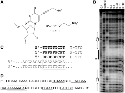
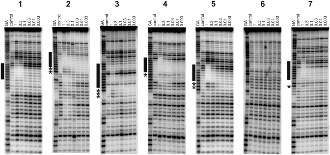
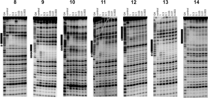
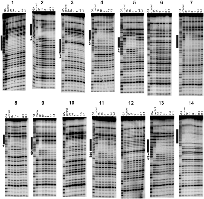
In order to enhance DNA triple helix stability synthetic oligonucleotides have been developed that bear amino groups on the sugar or base. One of the most effective of these is bis-amino-U (B), which possesses 5-propargylamino and 2'-aminoethoxy modifications. Inclusion of this modified nucleotide not only greatly enhances triplex stability, but also increases the affinity for related sequences. We have used a restriction enzyme protection, selection and amplification assay (REPSA) to isolate sequences that are bound by the heavily modified 9-mer triplex-forming oligonucleotide B(6)CBT. The isolated sequences contain A(n) tracts (n = 6), suggesting that the 5'-end of this TFO was responsible for successful triplex formation. DNase I footprinting with these sequences confirmed triple helix formation at these secondary targets and demonstrated no interaction with similar oligonucleotides containing T or 5-propargylamino-dU.
Figures
Similar articles
-
Stable DNA triple helix formation using oligonucleotides containing 2'-aminoethoxy,5-propargylamino-U.Biochemistry. 2002 Jun 11;41(23):7224-31. doi: 10.1021/bi020164n. Biochemistry. 2002. PMID: 12044153
-
DNA triple helix formation at target sites containing several pyrimidine interruptions: stabilization by protonated cytosine or 5-(1-propargylamino)dU.Biochemistry. 1999 Oct 12;38(41):13747-58. doi: 10.1021/bi9911637. Biochemistry. 1999. PMID: 10521282
-
Combining nucleoside analogues to achieve recognition of oligopurine tracts by triplex-forming oligonucleotides at physiological pH.FEBS Lett. 2005 Dec 5;579(29):6616-20. doi: 10.1016/j.febslet.2005.10.056. Epub 2005 Nov 9. FEBS Lett. 2005. PMID: 16293248
-
Selectivity and affinity of triplex-forming oligonucleotides containing 2'-aminoethoxy-5-(3-aminoprop-1-ynyl)uridine for recognizing AT base pairs in duplex DNA.Nucleic Acids Res. 2004 Aug 18;32(15):4439-47. doi: 10.1093/nar/gkh776. Print 2004. Nucleic Acids Res. 2004. PMID: 15317869 Free PMC article.
-
The anti-gene strategy: control of gene expression by triplex-forming-oligonucleotides.Anticancer Drug Des. 1991 Dec;6(6):569-84. Anticancer Drug Des. 1991. PMID: 1772570 Review.
Cited by
-
Synthesis and triplex-forming properties of oligonucleotides capable of recognizing corresponding DNA duplexes containing four base pairs.Nucleic Acids Res. 2015 Jul 13;43(12):5675-86. doi: 10.1093/nar/gkv496. Epub 2015 May 26. Nucleic Acids Res. 2015. PMID: 26013815 Free PMC article.
-
DNA Structural Changes Induced by Intermolecular Triple Helix Formation.ACS Omega. 2020 Jan 15;5(3):1679-1687. doi: 10.1021/acsomega.9b03776. eCollection 2020 Jan 28. ACS Omega. 2020. PMID: 32010842 Free PMC article.
-
Efficient access to 3'-terminal azide-modified RNA for inverse click-labeling patterns.Bioconjug Chem. 2014 Jan 15;25(1):188-95. doi: 10.1021/bc400513z. Epub 2013 Dec 20. Bioconjug Chem. 2014. PMID: 24358989 Free PMC article.
References
-
- Felsenfeld G, Davies DR, Rich A. Formation of a three-stranded polynucleotide molecule. J. Am. Chem. Soc. 1957;79:2023–2024.
-
- Fox KR. Targeting DNA with triplexes. Curr. Med. Chem. 2000;7:17–37. - PubMed
-
- Thuong NT, Hélène C. Sequence-specific recognition and modification of double-helical DNA by oligonucleotides. Angew. Chem. Intl. Ed. Engl. 1993;32:666–690.
-
- Doan TL, Perrouault L, Praseuth D, Habhoub N, Decout JL, Thuong NT, Lhomme J, Hélène C. Sequence-specific recognition, photo-cross-linking and cleavage of the DNA double helix by an oligo-[alpha]-thymidylate covalently linked to an azidoproflavine derivative. Nucleic Acids Res. 1987;15:7749–7760. - PMC - PubMed
-
- Moser HE, Dervan PB. Sequence-specific cleavage of double helical DNA by triple helix formation. Science. 1987;238:645–650. - PubMed
Publication types
MeSH terms
Substances
Grants and funding
LinkOut - more resources
Full Text Sources
Molecular Biology Databases
Miscellaneous
