Evolution of activation patterns during long-duration ventricular fibrillation in pigs
- PMID: 22180655
- PMCID: PMC3322740
- DOI: 10.1152/ajpheart.00419.2011
Evolution of activation patterns during long-duration ventricular fibrillation in pigs
Abstract
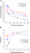
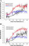
Quantitative analysis has demonstrated five temporal stages of activation during the first 10 min of ventricular fibrillation (VF) in dogs. To determine whether these stages exist in another species, we applied the same analysis to the first 10 min of VF recorded in vivo from two 504-electrode arrays, one each on left anterior and posterior ventricular epicardium in six anesthetized pigs. The following descriptors were continuously quantified: 1) number of wavefronts, 2) wavefront fractionations, 3) wavefront collisions, 4) repeatability, 5) multiplicity index, 6) wavefront conduction velocity, 7) activation rate, 8) mean area activated by the wavefronts, 9) negative peak rate of voltage change, 10) incidence of breakthrough/foci, 11) incidence of block, and 12) incidence of reentry. Cluster analysis of these descriptors divided VF into four stages (stages i-iv). The values of most descriptors increased during stage i (1-22 s after VF induction), changed quickly to values indicating greater organization during stage ii (23-39 s), decreased steadily during stage iii (40-187 s), and remained relatively unchanged during stage iv (188-600 s). The epicardium still activated during stage iv instead of becoming silent as in dogs. In conclusion, during the first 10 min, VF activation can be divided into four stages in pigs instead of five stages as in dogs. Following a 16-s period during the first minute of VF when activation became more organized, all parameters exhibited progressive decreased organization. Further studies are warranted to determine whether these changes, particularly the increased organization of stage ii, have clinical consequences, such as alteration in defibrillation efficacy.
Figures


References
-
- Antzelevitch C, Sicouri S, Litovsky SH, Lukas A, Krishnan SC, Di Diego JM, Gintant GA, Liu DW. Heterogeneity within the ventricular wall. Electrophysiology and pharmacology of epicardial, endocardial, and M cells. Circ Res 69: 1427–1449, 1991 - PubMed
-
- Bagdonas AA, Stuckey JH, Piera J, Amer NS, Hoffman BF. Effects of ischemia and hypoxia on the specialized conducting system of the canine heart. Am Heart J 61: 206–218, 1961 - PubMed
-
- Carmeliet E. Action potential duration, rate of stimulation, and intracellular sodium. J Cardiovasc Electrophysiol 17, Suppl 1: S2–S7, 2006 - PubMed
-
- Cobb LA, Fahrenbruch CE, Olsufka M, Copass MK. Changing incidence of out-of-hospital ventricular fibrillation, 1980–2000. JAMA 288: 3008–3013, 2002 - PubMed
-
- Cordeiro JM, Mazza M, Goodrow R, Ulahannan N, Antzelevitch C, Di Diego JM. Functionally distinct sodium channels in ventricular epicardial and endocardial cells contribute to a greater sensitivity of the epicardium to electrical depression. Am J Physiol Heart Circ Physiol 295: H154–H162, 2008 - PMC - PubMed
Publication types
MeSH terms
Grants and funding
LinkOut - more resources
Full Text Sources

