

Non-Fluent Speech in Frontotemporal Lobar Degeneration
- PMID: 22180700
- PMCID: PMC3238501
- DOI: 10.1016/j.jneuroling.2008.12.001
Non-Fluent Speech in Frontotemporal Lobar Degeneration
Abstract
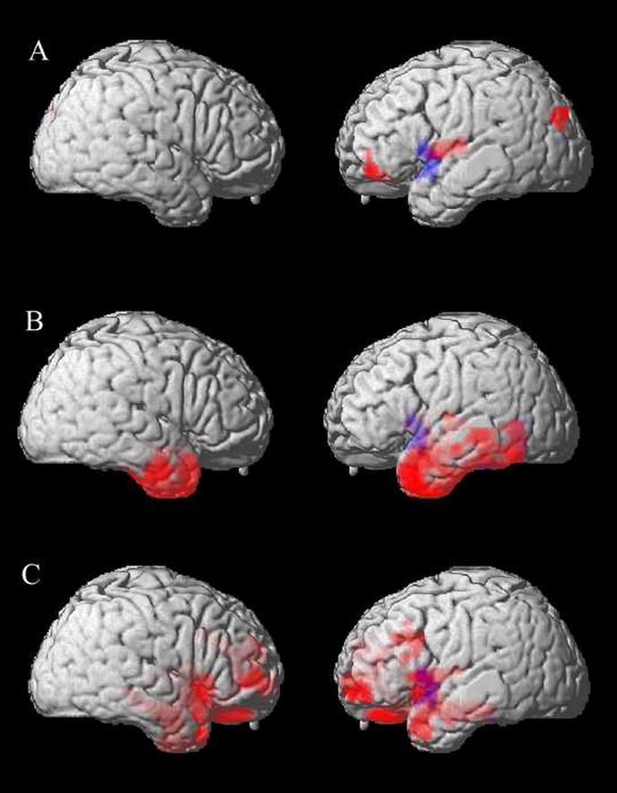
We investigated the cognitive and neural bases of impaired speech fluency, a central feature of primary progressive aphasia. Speech fluency was assessed in 35 patients with frontotemporal lobar degeneration (FTLD) who presented with progressive non-fluent aphasia (PNFA, n=11), semantic dementia (SemD, n=12), or a social and executive disorder without aphasia (SOC/EXEC, n=12). Fluency was quantified as the number of words per minute in an extended, semi-structured speech sample. This was related to language characteristics of the speech sample and to neuropsychological measures. PNFA patients were significantly less fluent than controls and other FTLD patients. Fluency correlated with grammatical expression but not with speech errors or executive difficulty. SemD and SOC/EXEC patients were also less fluent than controls. In SemD, fluency was associated with semantically limited content. In SOC/EXEC, fluency was associated with executive limitations. Voxel-based morphometry analyses of high-resolution MRI related fluency to gray matter volume in left inferior frontal, insula, and superior temporal regions for the entire cohort of FTLD patients. This region overlapped partially distinct atrophic areas in each FTLD subgroup. It thus appears to play a crucial role in speech fluency, which can be interrupted in different ways in different FTLD subgroups.
Figures
References
-
- Alexander MP, Benson DF, Stuss DT. Frontal lobes and language. Brain and Language. 1989;37:656–691. - PubMed
-
- Alexander MP, Naeser MA, Palumbo C. Broca’s area aphasia: aphasia after lesions including the frontal operculum. Neurology. 1990;40:353–362. - PubMed
-
- Amici S, Ogar J, Brambati SM, Miller BL, Neuhaus J, Dronkers NL, et al. Performance in specific language tasks correlates with regional volume changes in progressive aphasia. Cogn Behav Neurol. 2007;20(4):203–211. - PubMed
-
- Ash S, Moore P, Antani S, McCawley G, Work M, Grossman M. Trying to tell a tale: Discourse impairments in progressive aphasia and frontotemporal dementia. Neurology. 2006;66:1405–1413. - PubMed
Grants and funding
LinkOut - more resources
Full Text Sources