. 2011 Dec 12:2:250.
doi: 10.3389/fmicb.2011.00250. eCollection 2011.

Structural Basis for the Antiviral Activity of BST-2/Tetherin and Its Viral Antagonism

Affiliations

Structural Basis for the Antiviral Activity of BST-2/Tetherin and Its Viral Antagonism

Juan F Arias et al. Front Microbiol. .

Abstract

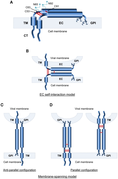
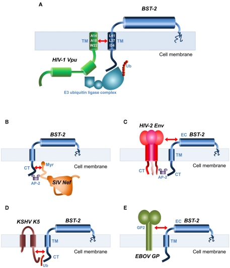
The interferon-inducible host restriction factor bone marrow stromal antigen 2 (BST-2/tetherin) blocks the release of HIV-1 and other enveloped viruses. In turn, these viruses have evolved specific antagonists to counteract this host antiviral molecule, such as the HIV-1 protein Vpu. BST-2 is a type II transmembrane protein with an unusual topology consisting of an N-terminal cytoplasmic tail (CT) followed by a single transmembrane (TM) domain, a coiled-coil extracellular (EC) domain, and a glycosylphosphatidylinositol (GPI) anchor at the C terminus. We and others showed that BST-2 restricts enveloped virus release by bridging the host and virion membranes with its two opposing membrane anchors and that deletion of either one completely abrogates antiviral activity. The EC domain also shows conserved structural properties that are required for antiviral function. It contains several destabilizing amino acids that confer the molecule with conformational flexibility to sustain the protein's function as a virion tether, and three conserved cysteine residues that mediate homodimerization of BST-2, as well as acting as a molecular ruler that separates the membrane anchors. Conversely, the efficient release of virions is promoted by the HIV-1 Vpu protein and other viral antagonists. Our group and others provided evidence from mutational analyses indicating that Vpu antagonism of BST-2-mediated viral restriction requires a highly specific interaction of their mutual TM domains. This interpretation is further supported and expanded by the findings of the latest structural modeling studies showing that critical amino acids in a conserved helical face of these TM domains are required for Vpu-BST-2 interaction and antagonism. In this review, we summarize the current advances in our understanding of the structural basis for BST-2 antiviral function as well as BST-2-specific viral antagonism.

Keywords: BST-2; HIV-1; Vpu; antagonist; interaction; restriction factor; transmembrane.


Figures

Figure 1
Figure 1
Topological characteristics of human BST-2. (A) Schematic representation of the domain structure of BST-2, a type II transmembrane (TM) protein. BST-2 features a short cytoplasmic N-terminus followed by an α-helical single-pass TM domain and an extended coiled-coil extracellular domain that is linked back to the plasma membrane by a C-terminal GPI anchor. N-glycosylation sites and cysteine residues for disulfide-bond formation in the extracellular domain (EC) are noted. (B–D) Topological models of BST-2’s functional state. (B) The EC self-interaction model, in which individual BST-2 monomers are anchored at both ends to the same membrane, with interaction between the ECs of cell-bound and virion-bound monomers. (C) Membrane-spanning anti-parallel model. Monomers are anchored in both membranes with opposing orientations. (D) Membrane-spanning parallel model. Monomers are anchored in both membranes with the same orientation.
Figure 2
Figure 2
Viral antagonists of BST-2 and their domains of interaction. Schematic representation of BST-2 and its known antagonists. The structural domains of interaction are indicated by red arrows. (A) HIV-1 Vpu and BST-2 interact through their mutual transmembrane (TM) domains. Key amino acid residues involved in the interaction are depicted in the TM helices. Also shown is the E3 ubiquitin (Ub) ligase complex required for BST-2 internalization. (B) SIV Nef recognizes the cytoplasmic (CT) domain of BST-2. The AP-2 clathrin adaptor recruited for BST-2 internalization is also shown. Myr, myristoylation site. (C) The envelope glycoprotein (Env) of HIV-2 and SIVtan binds to BST-2 through their mutual ectodomains (EC), and recruitment of AP-2 by the CT domain of Env required for internalization is also shown. (D) Kaposi’s sarcoma-associated herpesvirus (KSHV) K5 protein that is an ubiquitin ligase ubiquitinates a target lysine motif in the CT domain of BST-2, resulting in its internalization. (E) The antagonistic mechanisms of the Ebola virus (EBOV) glycoprotein (GP) are unclear, but require interaction between GP2 subunit of EBOV–GP and BST-2 EC.

References

    1. Abada P., Noble B., Cannon P. M. (2005). Functional domains within the human immunodeficiency virus type 2 envelope protein required to enhance virus production. J. Virol. 79, 3627–363810.1128/JVI.79.6.3627-3638.2005 - DOI - PMC - PubMed
    1. Aloia R. C., Tian H., Jensen F. C. (1993). Lipid composition and fluidity of the human immunodeficiency virus envelope and host cell plasma membranes. Proc. Natl. Acad. Sci. U.S.A. 90, 5181–518510.1073/pnas.90.11.5181 - DOI - PMC - PubMed
    1. Andrew A. J., Miyagi E., Kao S., Strebel K. (2009). The formation of cysteine-linked dimers of BST-2/tetherin is important for inhibition of HIV-1 virus release but not for sensitivity to Vpu. Retrovirology 6, 80.10.1186/1742-4690-6-S3-P80 - DOI - PMC - PubMed
    1. Andrew A. J., Miyagi E., Strebel K. (2011). Differential effects of human immunodeficiency virus type 1 Vpu on the stability of BST-2/tetherin. J. Virol. 85, 2611–261910.1128/JVI.02080-10 - DOI - PMC - PubMed
    1. Arnaud F., Black S. G., Murphy L., Griffiths D. J., Neil S. J., Spencer T. E., Palmarini M. (2010). Interplay between ovine bone marrow stromal cell antigen 2/tetherin and endogenous retroviruses. J. Virol. 84, 4415–442510.1128/JVI.00029-10 - DOI - PMC - PubMed

LinkOut - more resources