. 2011;6(12):e28501.
doi: 10.1371/journal.pone.0028501. Epub 2011 Dec 13.

Chymase-dependent generation of angiotensin II from angiotensin-(1-12) in human atrial tissue

Affiliations

Chymase-dependent generation of angiotensin II from angiotensin-(1-12) in human atrial tissue

Sarfaraz Ahmad et al. PLoS One. 2011.

Abstract

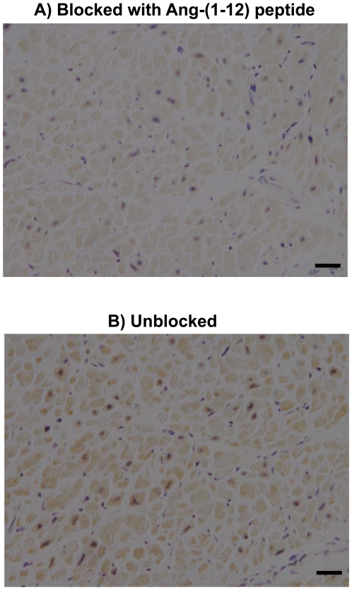
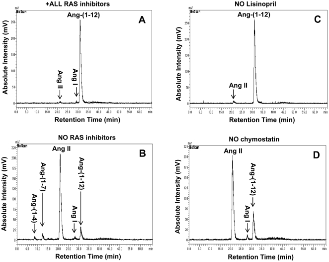
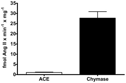
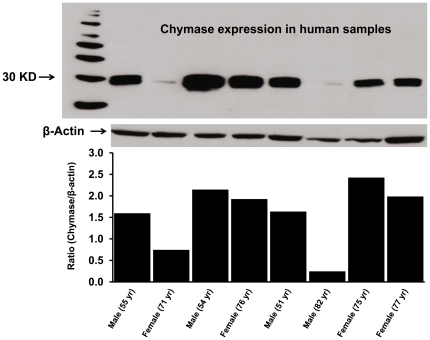
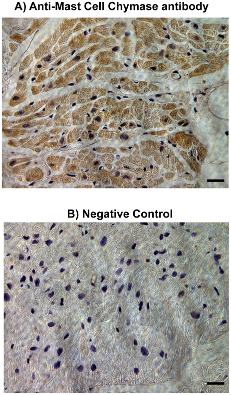
Since angiotensin-(1-12) [Ang-(1-12)] is a non-renin dependent alternate precursor for the generation of cardiac Ang peptides in rat tissue, we investigated the metabolism of Ang-(1-12) by plasma membranes (PM) isolated from human atrial appendage tissue from nine patients undergoing cardiac surgery for primary control of atrial fibrillation (MAZE surgical procedure). PM was incubated with highly purified ¹²⁵I-Ang-(1-12) at 37°C for 1 h with or without renin-angiotensin system (RAS) inhibitors [lisinopril for angiotensin converting enzyme (ACE), SCH39370 for neprilysin (NEP), MLN-4760 for ACE2 and chymostatin for chymase; 50 µM each]. ¹²⁵I-Ang peptide fractions were identified by HPLC coupled to an inline γ-detector. In the absence of all RAS inhibitor, ¹²⁵I-Ang-(1-12) was converted into Ang I (2±2%), Ang II (69±21%), Ang-(1-7) (5±2%), and Ang-(1-4) (2±1%). In the absence of all RAS inhibitor, only 22±10% of ¹²⁵I-Ang-(1-12) was unmetabolized, whereas, in the presence of the all RAS inhibitors, 98±7% of ¹²⁵I-Ang-(1-12) remained intact. The relative contribution of selective inhibition of ACE and chymase enzyme showed that ¹²⁵I-Ang-(1-12) was primarily converted into Ang II (65±18%) by chymase while its hydrolysis into Ang II by ACE was significantly lower or undetectable. The activity of individual enzyme was calculated based on the amount of Ang II formation. These results showed very high chymase-mediated Ang II formation (28±3.1 fmol × min⁻¹ × mg⁻¹, n = 9) from ¹²⁵I-Ang-(1-12) and very low or undetectable Ang II formation by ACE (1.1±0.2 fmol×min⁻¹ × mg⁻¹). Paralleling these findings, these tissues showed significant content of chymase protein that by immunocytochemistry were primarily localized in atrial cardiac myocytes. In conclusion, we demonstrate for the first time in human cardiac tissue a dominant role of cardiac chymase in the formation of Ang II from Ang-(1-12).


Conflict of interest statement

Competing Interests: The authors have declared that no competing interests exist.

Figures

Figure 1
Figure 1. Localization of Ang-(1-12) in human atrial tissue.
Comparative adjacent sections of Ang-(1-12) immunoreactivity obtained from human atrial tissue with protein A purified polyclonal antibody produced by AnaSpec. A) Antibody (1∶2,000 dilution) blocked with 100 µmol/L of human Ang-(1-12) peptide, and B) Unblocked antibody (1∶2,000 dilution). (Magnification 400; scale bar is 50 µm).
Figure 2
Figure 2. Ang-(1-12) metabolism by human atrial tissue.
The metabolism of 125I-Ang-(1-12) by plasma membrane isolated from human atrial tissues was analyzed by HPLC coupled to an inline BioScan γ-detector. The 125I-Ang-(1-12) was incubated with human plasma membrane for 60 min at 37°C with or without the inhibitor cocktail and the metabolites were separated by HPLC. A: Chromatograms represent the hydrolysis of 125I-Ang-(1-12) in the presence of all RAS inhibitors (All RAS inhibitor group containing lisinopril, SCH39370, MLN-4760, chymostatin, bestatin, amastatin, benzyl succinate, and PCMB). B: Hydrolysis of 125I-Ang-(1-12) in the absence of all RAS inhibitors cocktail (No RAS inhibitors group containing only bestatin, amastatin, benzyl succinate, and PCMB). C: Hydrolysis of 125I-Ang-(1-12) in the presence of the inhibitor cocktail that lacks only Lisinopril (No lisinopril inhibitor group containing all inhibitors as described in “A” except ACE inhibitor). D: Hydrolysis of 125I-Ang-(1-12) in the presence of inhibitor cocktail that lacks only chymostatin (No chymostatin group containing all inhibitors as described in “A” except chymase inhibitor). Before adding the 125I-Ang-(1-12), the plasma membrane was pre-incubated with inhibitors (each added at a dose of 50 µM) for 15 min at 37°C. The arrow indicates the retention time of 125I-Ang-(1-12) and its metabolic products. HPLC results are representative of three or more separate metabolism experiments for each human sample.
Figure 3
Figure 3. Chymase and ACE contribution to generate Ang II from Ang-(1-12).
Chymase and ACE enzyme mediated generation of Ang II product from the metabolism of 125I-Ang-(1-12) (1 nmol/L) by plasma membrane isolated from human atrial tissues. The contribution of each enzyme (chymase, and ACE) activity (fmol Ang II formation×min−1×mg−1) were calculated based on Ang II product analysis by HPLC in each human samples incubated with or without the chymostatin and lisinopril (each added at a dose 50 µM) for 60 min at 37°C. Data are expressed as mean ± SEM; total n = 9 (4 female).
Figure 4
Figure 4. Chymase protein expression in human atrial tissue.
Chymase protein expression in human atrial samples was analyzed by western blotting using a primary monoclonal anti-human chymase antibody (CMA1 antibody from R&D System, Cat# MAB-4099; 2 µg/mL). The plasma membrane (50 µg protein) were separated by gel electrophoresis and transferred on PVDF. Equal protein loading on each lane was confirmed by β-actin detection (1∶5,000 dilution). Level of protein expression was shown as relative O.D. ratio (chymase/β-actin) for each human sample.
Figure 5
Figure 5. Immunohistochemistry of human atrial tissue for chymase.
Immunostaining of human atrial tissue using an Anti-Mast Cell chymase antibody (Abcam Inc., Cambridge, MA; Cat# ab2377) revealed high expression of chymase within atrial cardiac myocytes (A). Negative control without primary antibody shows no staining for chymase (B). (Magnification 400; scale bar is 50 µm).

References

    1. Ferrario CM. New physiological concepts of the renin-angiotensin system from the investigation of precursors and products of angiotensin I metabolism. Hypertension. 2010;55:445–452. - PMC - PubMed
    1. Paul M, Poyan MA, Kreutz R. Physiology of local renin-angiotensin systems. Physiol Rev. 2006;86:747–803. - PubMed
    1. Nagata S, Kato J, Sasaki K, Minamino N, Eto T, et al. Isolation and identification of proangiotensin-12, a possible component of the renin-angiotensin system. Biochem Biophys Res Commun. 2006;350:1026–1031. - PubMed
    1. Prosser HC, Forster ME, Richards AM, Pemberton CJ. Cardiac chymase converts rat proAngiotensin-12 (PA12) to angiotensin II: effects of PA12 upon cardiac haemodynamics. Cardiovasc Res. 2009;82:40–50. - PubMed
    1. Prosser HC, Richards AM, Forster ME, Pemberton CJ. Regional vascular response to ProAngiotensin-12 (PA12) through the rat arterial system. Peptides. 2010;31:1540–1545. - PubMed

Publication types

MeSH terms