

Tracing of the Bile-chemotactic migration of juvenile Clonorchis sinensis in rabbits by PET-CT
- PMID: 22180795
- PMCID: PMC3236719
- DOI: 10.1371/journal.pntd.0001414
Tracing of the Bile-chemotactic migration of juvenile Clonorchis sinensis in rabbits by PET-CT
Abstract
Background: Adult Clonorchis sinensis live in the bile duct and cause clonorchiasis. It is known that the C. sinensis metacercariae excyst in the duodenum and migrate up to the bile duct through the common bile duct. However, no direct evidence is available on the in vivo migration of newly excysted C. sinensis juveniles (CsNEJs). Advanced imaging technologies now allow the in vivo migration and localization to be visualized. In the present study, we sought to determine how sensitively CsNEJs respond to bile and how fast they migrate to the intrahepatic bile duct using PET-CT.
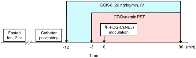
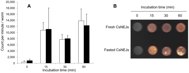
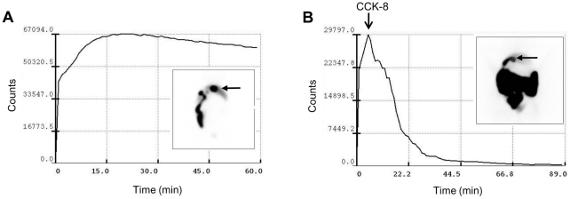
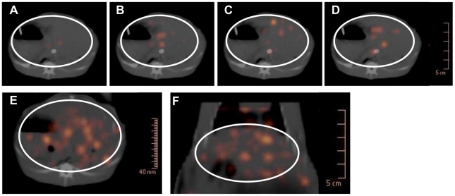
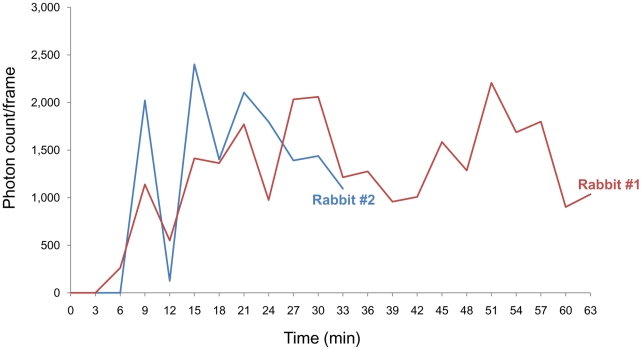
Methodology/principal findings: CsNEJs were radiolabeled with (18)F-fluorodeoxyglucose ((18)F-FDG). Rabbits with a gallbladder contraction response to cholecystokinin-8 (CCK-8) injection were pre-screened using cholescintigraphy. In these rabbits, gallbladders contracted by 50% in volume at an average of 11.5 min post-injection. The four rabbits examined were kept anesthetized and a catheter inserted into the mid duodenum. Gallbladder contraction was stimulated by injecting CCK-8 (20 ng/kg every minute) over the experiment. Anatomical images were acquired by CT initially and dynamic PET was then carried out for 90 min with a 3-min acquisition per frame. Twelve minutes after CCK-8 injection, about 3,000 (18)F-FDG-labeled CsNEJs were inoculated into the mid duodenum through the catheter. Photon signals were detected in the liver 7-9 min after CsNEJs inoculation, and these then increased in the whole liver with stronger intensity in the central area, presenting that the CsNEJs were arriving at the intrahepatic bile ducts.
Conclusion: In the duodenum, CsNEJs immediately sense bile and migrate quickly with bile-chemotaxis to reach the intrahepatic bile ducts by way of the ampulla of Vater.
Conflict of interest statement
The authors have declared that no competing interests exist.
Figures
References
-
- Hong ST, Fang Y. Clonorchis sinensis and clonorchiasis, an update. Parasitol Int. 2011 D.O.I.: 10.1016/j.parint.2011.06.007. - PubMed
-
- Rim HJ. The current pathobiology and chemotherapy of clonorchiasis. Korean J Parasitol. 1986;24(Suppl):1–141. - PubMed
-
- Rim HJ. Clonorchiasis: an update. J Helminthol. 2005;79:269–281. - PubMed
-
- Bouvard V, Baan R, Straif K, Grosse Y, Secretan B, et al. A review of human carcinogens-Part B: biological agents. Lancet Oncol. 2009;10:321–322. - PubMed
