

Microencapsulating and Banking Living Cells for Cell-Based Medicine
- PMID: 22180835
- PMCID: PMC3237390
- DOI: 10.1260/2040-2295.2.4.427
Microencapsulating and Banking Living Cells for Cell-Based Medicine
Abstract
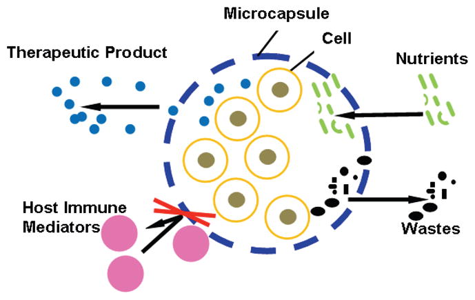
A major challenge to the eventual success of the emerging cell-based medicine such as tissue engineering, regenerative medicine, and cell transplantation is the limited availability of the desired cell sources. This challenge can be addressed by cell microencapsulation to overcome the undesired immune response (i.e., to achieve immunoisolation) so that non-autologous cells can be used to treat human diseases, and by cell/tissue preservation to bank living cells for wide distribution to end users so that they are readily available when needed in the future. This review summarizes the status quo of research in both cell microencapsulation and banking the microencapsulated cells. It is concluded with a brief outlook of future research directions in this important field.
Conflict of interest statement
Figures
References
-
- Chang TM. Semipermeable Microcapsules. Science. 1964;146:524–525. - PubMed
-
- Lim F, Sun AM. Microencapsulated Islets as Bioartificial Endocrine Pancreas. Science. 1980;210(4472):908–910. - PubMed
-
- Murua A, Portero A, Orive G, Hernandez RM, de Castro M, Pedraz JL. Cell microencapsulation technology: towards clinical application. J Control Release. 2008;132(2):76–83. - PubMed
-
- Orive G, Hernandez RM, Gascon AR, Calafiore R, Chang TM, De Vos P, Hortelano G, Hunkeler D, Lacik I, Shapiro AM, Pedraz JL. Cell encapsulation: promise and progress. Nat Med. 2003;9(1):104–107. - PubMed
-
- Orive G, Hernandez RM, Rodriguez Gascon A, Calafiore R, Chang TM, de Vos P, Hortelano G, Hunkeler D, Lacik I, Pedraz JL. History, challenges and perspectives of cell microencapsulation. Trends Biotechnol. 2004;22(2):87–92. - PubMed
Grants and funding
LinkOut - more resources
Full Text Sources
Other Literature Sources
Molecular Biology Databases
