

Bone loss following spinal cord injury in a rat model
- PMID: 22181016
- PMCID: PMC3353757
- DOI: 10.1089/neu.2011.2037
Bone loss following spinal cord injury in a rat model
Abstract
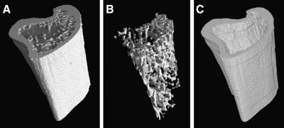
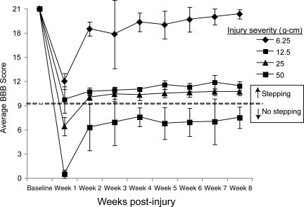
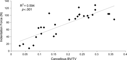
The current study was undertaken to follow the time course of bone loss in the proximal tibia of rats over several weeks following thoracic contusion spinal cord injury (SCI) of varying severity. It was hypothesized that bone loss would be more pronounced in the more severely injured animals, and that hindlimb weight bearing would help prevent bone loss. Twenty-six female Sprague-Dawley rats (200-225 g, 6-7 weeks old) received standard thoracic (T9) injuries at energies of 6.25, 12.5, 25, or 50 g-cm. The rats were scored weekly for hindlimb function during locomotion. At 0, 2 or 3, and 8 weeks, high-resolution micro-CT images of each right tibia were obtained. Mechanical indentation testing was done to measure the compressive strength of the cancellous bone structure. The 6.25 g-cm group showed near normal locomotion, the 12.5 and 25 g-cm groups showed the ability to frequently or occasionally generate weight-supported plantar steps, respectively, and the 50 g-cm group showed only movement without weight-supported plantar stepping. The 6.25, 12.5 and 25 g-cm groups remained at the same level of bone volume fraction (cancBV/TV=0.24±0.07), while the 50 g-cm group experienced severe bone loss (67%), resulting in significantly lower (p<0.05) bone volume fraction (cancBV/TV=0.11±0.05) at 8 weeks. Proximal tibia cancellous bone strength was reduced by approximately 50% in these severely injured rats. Instead of a linear proportionality between injury severity and bone loss, there appears to be a distinct functional threshold, marked by occasional weight-supported stepping, above which bone loss does not occur.
Figures


Similar articles
-
Changes in the structural and material properties of the tibia in patients with spinal cord injury.Spinal Cord. 2012 Apr;50(4):333-7. doi: 10.1038/sc.2011.143. Epub 2011 Nov 29. Spinal Cord. 2012. PMID: 22124349
-
Spinal cord injury causes more damage to bone mass, bone structure, biomechanical properties and bone metabolism than sciatic neurectomy in young rats.Osteoporos Int. 2006 Oct;17(10):1552-61. doi: 10.1007/s00198-006-0165-3. Epub 2006 Jul 28. Osteoporos Int. 2006. PMID: 16874443
-
Spinal cord injury causes rapid osteoclastic resorption and growth plate abnormalities in growing rats (SCI-induced bone loss in growing rats).Osteoporos Int. 2008 May;19(5):645-52. doi: 10.1007/s00198-007-0494-x. Epub 2007 Nov 7. Osteoporos Int. 2008. PMID: 17987335 Free PMC article.
-
Passive bicycle training stimulates epiphyseal bone formation and restores bone integrity independent of locomotor recovery in a rat spinal cord injury model.J Appl Physiol (1985). 2024 Sep 1;137(3):676-688. doi: 10.1152/japplphysiol.00299.2024. Epub 2024 Aug 1. J Appl Physiol (1985). 2024. PMID: 39088645
-
Bone loss at the distal femur and proximal tibia in persons with spinal cord injury: imaging approaches, risk of fracture, and potential treatment options.Osteoporos Int. 2017 Mar;28(3):747-765. doi: 10.1007/s00198-016-3798-x. Epub 2016 Dec 5. Osteoporos Int. 2017. PMID: 27921146 Review.
Cited by
-
Reactions of the rat musculoskeletal system to compressive spinal cord injury (SCI) and whole body vibration (WBV) therapy.J Musculoskelet Neuronal Interact. 2015 Jun;15(2):123-36. J Musculoskelet Neuronal Interact. 2015. PMID: 26032204 Free PMC article.
-
The Efficacy of Body-Weight Supported Treadmill Training and Neurotrophin-Releasing Scaffold in Minimizing Bone Loss Following Spinal Cord Injury.Bioengineering (Basel). 2024 Aug 12;11(8):819. doi: 10.3390/bioengineering11080819. Bioengineering (Basel). 2024. PMID: 39199776 Free PMC article.
-
Testosterone dose dependently prevents bone and muscle loss in rodents after spinal cord injury.J Neurotrauma. 2014 May 1;31(9):834-45. doi: 10.1089/neu.2013.3155. J Neurotrauma. 2014. PMID: 24378197 Free PMC article.
-
Rehabilitation: Neurogenic Bone Loss after Spinal Cord Injury.Biomedicines. 2023 Sep 20;11(9):2581. doi: 10.3390/biomedicines11092581. Biomedicines. 2023. PMID: 37761022 Free PMC article. Review.
-
Decrease of PPARδ in Type-1-Like Diabetic Rat for Higher Mortality after Spinal Cord Injury.PPAR Res. 2014;2014:456386. doi: 10.1155/2014/456386. Epub 2014 Apr 10. PPAR Res. 2014. PMID: 24817882 Free PMC article.
References
-
- Basso D.M. Beattie M.S. Bresnahan J.C. A sensitive and reliable locomotor rating scale for open field testing in rats. J. Neutrotrauma. 1995;12:1–21. - PubMed
-
- Basso D.M. Beattie M.S. Bresnahan J.C. Graded histological and locomotor outcomes after spinal cord contusion using the NYU weight-drop device versus transection. Exp. Neurol. 1996;139:244–256. - PubMed
-
- Biering-Sorensen F. Bohr H. Schaadt O. Bone mineral content of the lumbar spine and lower extremities years after spinal cord lesion. Paraplegia. 1988;26:293–301. - PubMed
-
- Biering-Sørensen F. Hansen B. Lee B.S. Non-pharmacological treatment and prevention of bone loss after spinal cord injury: a systematic review. Spinal Cord. 2009;47:508–518. - PubMed
Publication types
MeSH terms
Grants and funding
LinkOut - more resources
Full Text Sources
Medical
