Review
. 2012 Mar;31(2):136-51.
doi: 10.1016/j.preteyeres.2011.11.005. Epub 2011 Dec 10.

CNTF and retina

Affiliations
Review

CNTF and retina

Rong Wen et al. Prog Retin Eye Res. 2012 Mar.

Abstract

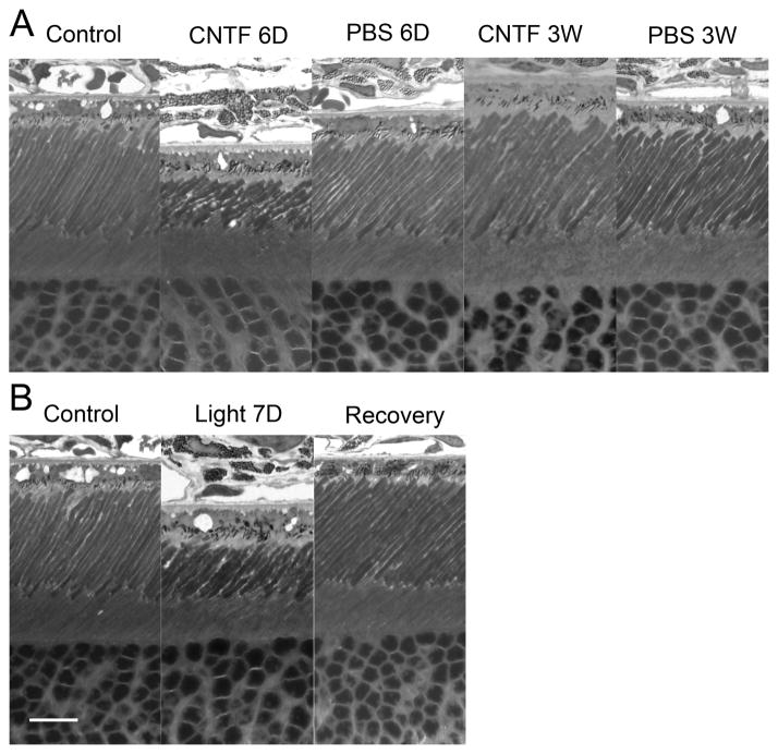
Ciliary neurotrophic factor (CNTF) is one of the most studied neurotrophic factors for neuroprotection of the retina. A large body of evidence demonstrates that CNTF promotes rod photoreceptor survival in almost all animal models. Recent studies indicate that CNTF also promotes cone photoreceptor survival and cone outer segment regeneration in the degenerating retina and improves cone function in dogs with congenital achromotopsia. In addition, CNTF is a neuroprotective factor and an axogenesis factor for retinal ganglion cells (RGCs). This review focuses on the effects of exogenous CNTF on photoreceptors and RGCs in the mammalian retina and the potential clinical application of CNTF for retinal degenerative diseases.


Figures

Fig. 1
Fig. 1
Schematic illustration of CNTF signaling through STAT3. CNTF binds to the receptor complex of CNTFRα, gp130, and LIFRβ and activates JAK kinase. Activated JAK kinase phosphorylates tyrosine residues (P) of the intracellular domain of gp130 and LIF, which provide docking sites for STAT3. After STAT3 is phosphorylated at the docking sites by JAK kinase, phospho-STAT3 (pSTAT3) forms dimers and translocates to the nucleus to induce gene transcription.
Fig. 2
Fig. 2
Protection of photoreceptors by CNTF. Eyes of hererozygous transgenic rats carrying the rhodopsin mutation S334ter (Liu et al., 1999) were intravitreally injected with either NTC-200 cells (parental cell line, n=6) or NTC-201 cells (CNTF secreting cell line, n=6) at postnatal day 9 (PD 9). Retinas were examined at PD 20. In the untreated (A) or NTC-200 cell (B) treated retinas, only 1–2 rows of photoreceptor nuclei remained in the outer nuclear layer (ONL, white brackets), whereas in the NTC-201 cell treated eyes, the ONL contained 5–6 row of nuclei (C). From Tao et al. (2000) (Copyright by the Association for Research in Vision and Ophthalmology).
Fig. 3
Fig. 3
Dark-adapted ERG responses in CNTF treated rat retina. ERG waveforms at increasing stimulus intensities from top to bottom recorded from retinas treated with PBS (right eye) or CNTF (10 μg, left eye) (A). Average amplitudes of the a-wave (B, C) and the b-wave (D, E) at each intensity at 6 days (B, D) and 21 days (C, E) after treatment. CNTF induced a significant decrease in the a- and b-wave 6 days after treatment. No significant difference between CNTF- and PBS-treated eyes in either a- or b-wave was seen 21 days after injection. Solid lines in B and C are B-spline curves through the data points. Solid lines in D and E are the averaged Naka-Rushton function fits. Dashed lines indicate Vmax and k values from these fits. Data points are averages of 8 animals and error bars are ± SEM. See the original paper (Wen, et al. 2006) for details. From Wen, et al. (2006) with permission.
Fig. 4
Fig. 4
Changes of ROS induced by CNTF and light exposure. A significant shortening of the ROS were seen in eyes 6 days after CNTF treatment (A). These changes were completely reversed 3 weeks after CNTF treatment (A). No alteration of ROS was observed in eyes treated with PBS (A). A shortening of ROS was observed in eyes after 7 days of light exposure (400 lux, 10hr daily) (B). The ROS length recovered to normal after 7 days in cyclic 50 lux light (B). Scale bar: 10 μm. Modified from Wen et al. (2008).
Fig. 5
Fig. 5
Schematic illustration of hypothesized mechanism of CNTF- and light-induced photoreceptor plasticity. Exogenous CNTF directly activates Müller cells, which in turn signal rod photoreceptors to down-regulate the phototransduction machinery. In photostasis, Müller cells collect information of the overall number of photons captured by photoreceptors along with circadian information from photosensitive ganglion cells. When the number of photons captured exceeds the daily quota for the retina, Müller cells inform rods to down regulate their phototransduction machinery. The CNTF and photostasis pathways converge in Müller cells. From Wen et al. (2008).
Fig. 6
Fig. 6
Regeneration of COS induced by CNTF treatment. Retinas (superior regions) of S334ter-3 rats at PD 35 were treated with either CNTF or PBS (phosphate-buffered saline) and harvested 10 days after treatment. Whole-mounted retinas of PD 35 (A, before treatment), PD 45 treated with PBS at PD 35 (B), and PD 45 treated with CNTF at PD 35 (C) were stained with PNA. More PNA-positive cells were seen in CNTF treated retinas than those in either PBS treated retinas or PD 35 untreated retinas. Quantitative analysis showed that PNA-positive cells were significantly more in the PD 45 CNTF treated retinas (657±76, n=4) than in the PD 35 retinas (467±73, n=3), or PD 45 retinas treated with PBS (334±33, n=3) (D, mean ± SD). Asterisk indicates P<0.05; double-asterisk indicates P ≤0.01 (ANOVA analysis and Bonferroni test). Scale bar: 100 μm. From Li et al. (2010).
Fig. 7
Fig. 7
CNTF-induced phosphorylation of STAT3. Immunostaining of phospho-STAT3 was mainly detected in the nuclei of ganglion cells in the untreated retina (A). CNTF induced a dramatic increase in STAT3 phosphorylation in a specific band of cells in the INL (B). Müller cells were identified by antibodies against GS (glutamine synthetase) (C, red). The immunoreactivities of pSTAT3 (green) and GS (red) are clearly co-localized (D, yellow). OS, outer segments; ONL, outer nuclear layer; INL, inner nuclear layer; GCL, ganglion cell layer. Scale bar: 50 μm. Modified from Wen et al. (2006) with permission.
Fig. 8
Fig. 8
Schematic illustration of CNTF-secreting implant using Encapsulated Cell Technology. The implant is composed of a section of semi-permeable membrane capsule which contains CNTF secreting cells and scaffold. The membrane capsule is sealed at both ends with a suture clip at one end for anchoring on the sclera (A). The membrane allows O2 and nutrients to diffuse in and therapeutic agent (CNTF in this case) to diffuse out. It also keeps components of the immune system out (B). The implant is 6 mm in length and 1 mm in diameter. It is outside the visual axis of the eye when anchored to the sclera (C).
Fig. 9
Fig. 9
Effect of intraocular CNTF on visual acuity stabilization. Fifty-one patients with geographic atrophy were randomly treated with high- or low-dose CNTF implants or sham operated. As measured at 12 months post implantation, 96.3% of the high-dose eyes lost <3 lines (<15 letters), whereas 75% of sham treated eyes lost <15 letters (P=0.078) (A). For patients with baseline vision at 20/63 or better, 100% in the high-dose group lost <15 letters whereas 55.6% in the combined low-dose/sham group lost < 15 letters, (P=0.033). Modified from Zhang et al. (2011).

References

    1. Adijanto J, Banzon T, Jalickee S, Wang NS, Miller SS. CO2-induced ion and fluid transport in human retinal pigment epithelium. J Gen Physiol. 2009;133:603–622. - PMC - PubMed
    1. Adler R. Ciliary neurotrophic factor as an injury factor. Curr Opin Neurobiol. 1993;3:785–789. - PubMed
    1. Adler R, Landa KB, Manthorpe M, Varon S. Cholinergic neuronotrophic factors: intraocular distribution of trophic activity for ciliary neurons. Science. 1979;204:1434–1436. - PubMed
    1. Barbin G, Manthorpe M, Varon S. Purification of the chick eye ciliary neuronotrophic factor. J Neurochem. 1984;43:1468–1478. - PubMed
    1. Bargoot FG, Williams TP, Beidler LM. The localization of radioactive amino acid taken up into the outer segments of frog (Rana pipiens) rods. Vision Res. 1969;9:385–391. - PubMed

Publication types

Substances

LinkOut - more resources