. 2012 Mar;212(3):327-42.
doi: 10.1530/JOE-11-0300. Epub 2011 Dec 19.

Chronic pulsatile hyperglycemia reduces insulin secretion and increases accumulation of reactive oxygen species in fetal sheep islets

Affiliations

Chronic pulsatile hyperglycemia reduces insulin secretion and increases accumulation of reactive oxygen species in fetal sheep islets

Alice S Green et al. J Endocrinol. 2012 Mar.

Abstract

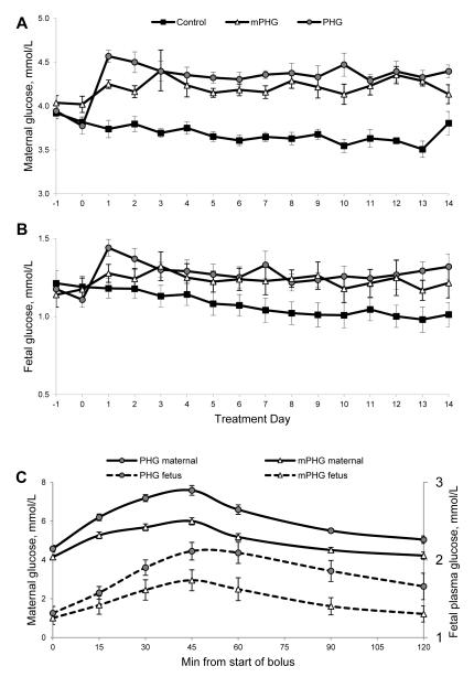
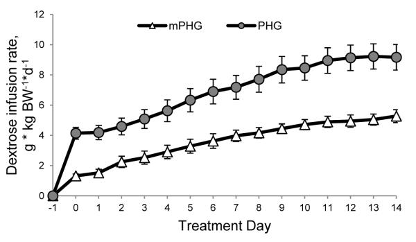
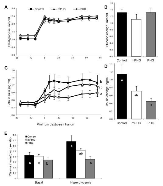
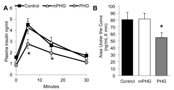
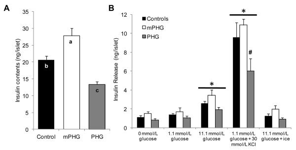
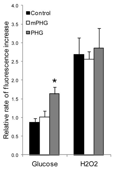
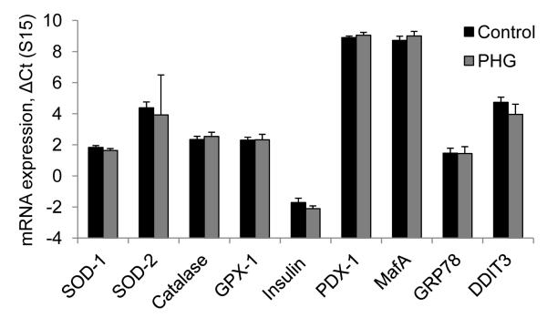
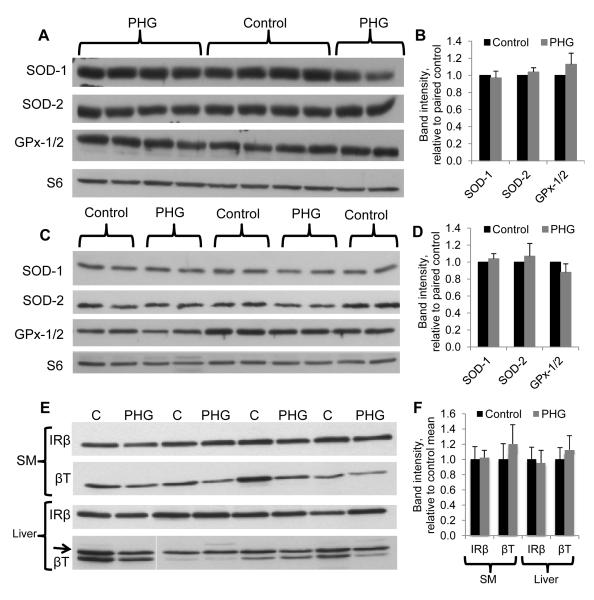
Children from diabetic pregnancies have a greater incidence of type 2 diabetes. Our objective was to determine if exposure to mild-moderate hyperglycemia, by modeling managed diabetic pregnancies, affects fetal β-cell function. In sheep fetuses, β-cell responsiveness was examined after 2 weeks of sustained hyperglycemia with 3 pulses/day, mimicking postprandial excursions, and compared to saline-infused controls (n = 10). Two pulsatile hyperglycemia (PHG) treatments were studied: mild (mPHG, n = 5) with +15% sustained and +55% pulse; and moderate (PHG, n = 10) with +20% sustained and +100% pulse. Fetal glucose-stimulated insulin secretion and glucose-potentiated arginine insulin secretion were lower (P < 0.05) in PHG (0.86 ± 0.13 and 2.91 ± 0.39 ng/ml plasma insulin) but not in mPHG fetuses (1.21 ± 0.08 and 4.25 ± 0.56 ng/ml) compared to controls (1.58 ± 0.25 and 4.51 ± 0.56 ng/ml). Islet insulin content was 35% lower in PHG and 35% higher in mPHG vs controls (P < 0.01). Insulin secretion and maximally stimulated insulin release were also reduced (P < 0.05) in PHG islets due to lower islet insulin content. Isolated PHG islets also had 63% greater (P < 0.01) reactive oxygen species (ROS) accumulation at 11.1 mmol/l glucose than controls (P < 0.01), but oxidative damage was not detected in islet proteins. PHG fetuses showed evidence of oxidative damage to skeletal muscle proteins (P < 0.05) but not insulin resistance. Our findings show that PHG induced dysregulation of islet ROS handling and decreased islet insulin content, but these outcomes are independent. The β-cell outcomes were dependent on the severity of hyperglycemia because mPHG fetuses had no distinguishable impairments in ROS handling or insulin secretion but greater insulin content.


Figures

Figure 1
Figure 1. Maternal (A) and Fetal (B) Plasma Glucose Concentrations
Sustained mild (mPHG) or moderate (PHG) hyperglycemia was initiated on day 0. Plasma was sampled prior to initiating treatment on day 0 and prior to the 0800 dextrose pulse daily thereafter. Panel C shows maternal and fetal plasma glucose concentrations during the 45-min dextrose pulse administered at 1400 on day 9 of treatment. The two-hour sampling period illustrates the magnitude and duration of hyperglycemia resulting from one of the dextrose pulses, which were given 3x/day throughout the treatment period.
Figure 2
Figure 2. Dextrose Infusion Rates Increase during mPHG and PHG Treatment Periods
Rates of dextrose infusion required to chronically maintain maternal plasma glucose 15% (mPHG) and 20% (PHG) above euglycemia increased during the course of the 14-day treatment.
Figure 3
Figure 3. Impaired Glucose Stimulated Insulin Secretion in PHG Fetuses
Graphs A and C show mean plasma glucose and insulin concentrations during basal (−21 to 0 minutes) and hyperglycemic (after time 0) periods. The bar graphs show the changes in glucose (B) and insulin (D) between the basal and hyperglycemic (45-61 minutes) steady state periods, which were calculated as the difference between hyperglycemia and basal mean concentrations. The elevation in glucose was similar between treatments, but PHG fetuses had a lower insulin response than control fetuses (P<0.05). Plasma insulin/glucose ratios (panel E) are shown for basal and hyperglycemic steady state periods. Bars not sharing the same letter are different, P<0.05.
Figure 4
Figure 4. Impaired GPAIS in PHG Fetuses
The follow-on arginine bolus administered at ~65 min into the square-wave hyperglycemic clamp stimulated insulin secretion in all treatment groups and is presented relative to the start of the arginine infusion in panel A. An interaction between treatment and sample time (P<0.05) was found, and insulin concentrations were lower (P<0.05) in PHG fetuses at 5 and 15 minutes, indicated by the asterisks. In panel B, GPAIS area under the curve (ng/mL X minute), calculated using basal steady state period insulin concentration as baseline, is reduced in PHG fetuses (* = P≤0.05).
Figure 5
Figure 5. Decreased Insulin Content and Release in PHG Islets
In panel A, mean insulin content (ng/islet) is presented for islets isolated from control (n=7), mPHG (n=5), and PHG (n=8) fetuses (indicated on the x-axis). In panel B, insulin release (ng/islet) is presented for static incubations in KRB/BSA media containing 0 mmol/L glucose, 1.1 mmol/L glucose, 11.1 mmol/L glucose, 30 mmol/L KCl and 1.1 mmol/L glucose, or 11.1 mmol/L glucose incubated on ice (indicated on the abscissa). Insulin release was increased in 11.1 mmol/L glucose and 30 mmol/L KCl compared to incubation with 0 and 1.1 mmol/L glucose, and is indicated by the horizontal bars with an asterisk (P<0.05). In the PHG islets, KCl stimulated insulin release was less than control and mPHG islets, which is indicated with the number symbol (P<0.05).
Figure 6
Figure 6. Greater Glucose Stimulated ROS Accumulation in PHG Islets
In isolated fetal sheep islets, the fluorescence intensities from oxidation of the CM-H2DCFDA probe were determined in 1.1 mmol/L glucose, 11.1 mmol/L glucose, and 9 mmol/L H2O2 every minute for 15 minutes in each condition. Rates of ROS accumulation in stimulatory glucose and H2O2 were normalized to the basal rate (1.1 mmol/L glucose) for each islet, and the bars represent the mean ± SEM for 2-5 islets from each of eight control, five mPHG, and six PHG fetuses. The asterisk indicates a significant difference (P<0.05) between control and PHG treatments in ROS accumulation at 11.1 mmol/L glucose.
Figure 7
Figure 7. Islet Gene Expression for Antioxidant Enzymes, Insulin, and ER Stress
The mRNA expression normalized to ribosomal protein s15 is shown for enzymatic antioxidant defense (SOD-1, SOD-2, catalase, and GPX-1); insulin transcription (insulin, PDX-1, and MafA); and endoplasmic reticulum stress (GRP78 and DDIT3) in islets isolated from control and PHG fetuses. Values shown are calculated by the comparative ΔCT method (CT gene of interest - CT reference gene). Gene expression was not different between control and PHG islets.
Figure 8
Figure 8. Antioxidant Enzyme and Insulin Receptor Concentrations in Liver and Skeletal Muscle
Representative Western immunoblots show protein concentrations of SOD-1, SOD-2, GPx-1/2, and the reference protein ribosomal protein S6, in skeletal muscle (A and B) and liver (C and D) and of insulin receptor β and the reference protein β-tubulin in skeletal muscle (SM) and liver (E and D). Bar graphs show quantification of band density by analysis in ImageJ. Antioxidant enzyme and insulin receptor β protein concentrations were not different between control and PHG treatments.

References

    1. Aerts L, Holemans K, Van Assche FA. Maternal diabetes during pregnancy: consequences for the offspring. Diabetes Metab Rev. 1990;6:147–167. - PubMed
    1. Aerts L, van Assche FA. Rat foetal endocrine pancreas in experimental diabetes. J Endocrinol. 1977;73:339–346. - PubMed
    1. Aerts L, Van Assche FA. Endocrine pancreas in the offspring of rats with experimentally induced diabetes. J Endocrinol. 1981;88:81–88. - PubMed
    1. Aerts L, Vercruysse L, Van Assche FA. The endocrine pancreas in virgin and pregnant offspring of diabetic pregnant rats. Diabetes Res Clin Pract. 1997;38:9–19. - PubMed
    1. American Diabetes Association Gestational Diabetes Mellitus. Diabetes Care. 2004;27:S88–90. - PubMed

Publication types