. 2012 Mar;47(3):175-82.
doi: 10.1097/RLI.0b013e318234e75b.

Compartmental analysis of renal BOLD MRI data: introduction and validation

Affiliations

Compartmental analysis of renal BOLD MRI data: introduction and validation

Behzad Ebrahimi et al. Invest Radiol. 2012 Mar.

Abstract

Objectives: Functional blood oxygenation level-dependent (BOLD) magnetic resonance imaging is a powerful tool to assess renal function, but BOLD analysis using T2* image differentiation of cortex and medulla is laborious and prone to errors. We developed and validated an alternative compartmental analysis method to differentiate renal cortical and medullary BOLD relaxivity index, R2*. This method uses whole-kidney regions of interest (ROI), thus eliminating the need for anatomic cortical and medullary definition.

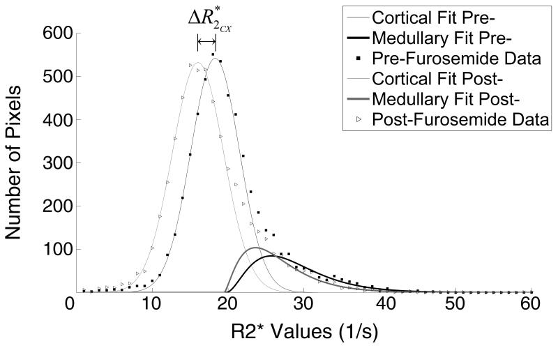
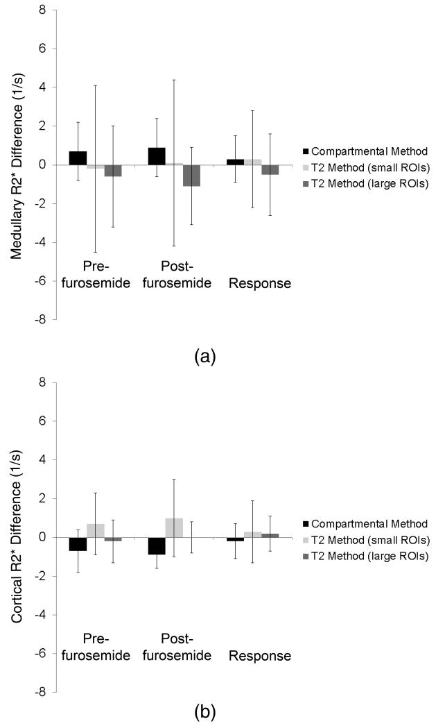
Materials and methods: Nine hypertensive patients and 11 domestic pigs, some with renal artery stenosis, were studied using BOLD MRI before and after injection of furosemide, which reduces medullary oxygen consumption. R2* in cortex and medulla estimated before and after furosemide with the compartmental method were compared with those obtained using conventional T2* image selection for ROI (manual ROI method), and a reference method with ROIs obtained using contrast-enhanced computerized tomography images were coregistered for the same kidneys.

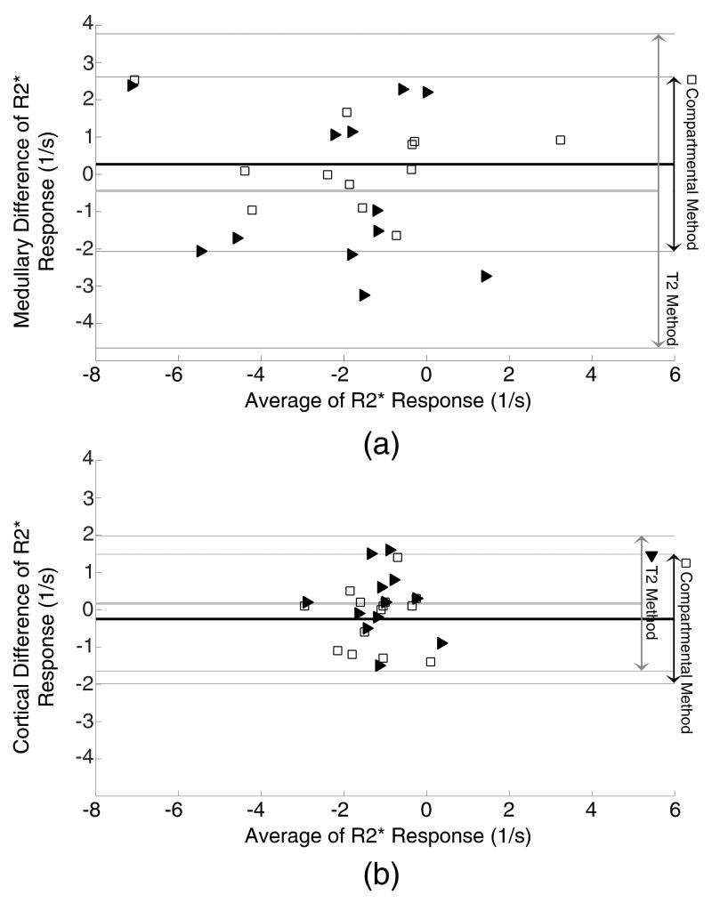
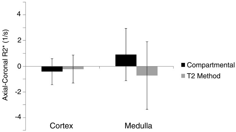
Results: All 3 methods provided similar cortical R2* values, but the Bland-Altman methods' agreement confidence intervals of the reference and compartmental-derived medullary R2* response in humans and pigs were smaller than those in the manual ROI method. Operator dependency in swine was lower in the compartmental method, and its estimates of variation were almost 1/3 compared with the manual ROI method.

Conclusions: The new compartmental method, which is less labor intensive than the conventional method, provides comparable and less variable kidney R2* estimations, especially in renal medulla. This method could be useful for analysis of kidney BOLD data.


Figures

Figure 1
Figure 1
ROI selection in the 4 methods studied. The small ROIs (a) and large ROIs (b) T2, the hybrid (c), and the compartmental (d) methods. In the hybrid method ROIs were selected to exclude potential overlap within the compartments to minimize volume averaging.
Figure 2
Figure 2
A contrast-enhanced MDCT image (a), a corresponding MRI parametric R2* map (b), and the resultant histogram showing the best fitted cortical and medullary curves (c). Region (1) shows the low R2* components, such as urine, and (2) shows high R2* components, such as highly hypoxic regions. The high R2* region in the vicinity of a low R2* regions on the parametric map is prone to mis-registration, while on the histogram the two regions are mapped to the two extreme ends of spectrum and have no mutual effect.
Figure 3
Figure 3
Examples of coronal and axial images constructed for the reference “hybrid” method of BOLD MR analysis. MDCT (a,d) and MR (c,f) images were co-registered (b,e) based on anatomical landmarks. Contrast agent aided MDCT images were used to select ROIs in the hybrid method. The quality of co-registration has been demonstrated with point-to-point verification of the edge of the kidney (+), medullary regions (o), and landmarks (x).
Figure 4
Figure 4
Representative cortical and medullary curves obtained before and after furosemide administration. Cortical curve shifted slightly left towards lower R2* values post-furosemide (ΔR2*), while the medullary curve showed both a shift and a change in the distribution pattern.
Figure 5
Figure 5
Altman-Bland graphs comparing the bias and variation of R2* derived from the compartmental (blank squares) and T2 (solid triangles) methods to the reference hybrid method. The biases in both methods were small, but the compartment method showed less variability (higher reproducibility).
Figure 6
Figure 6
R2* values in the pig medulla (top) and cortex (bottom) calculated using the compartmental and small and large-ROI T2 methods before and after furosemide, as well as the response intervention. Estimated cortical and medullary R2* values are close, but the compartmental method shows smaller variations in medullary values than the T2 method.
Figure 7
Figure 7
The variation of R2* values calculated from coronal and axial images of the same pig kidneys. The biases in both methods were small. However, the variation was larger in the T2 than in the compartmental method and in the medulla (a) than in the cortex (b).
Figure 8
Figure 8
R2* values in the human medulla (top) and cortex (bottom) calculated using the compartmental and T2 methods before and after furosemide, and the response to intervention, compared to the hybrid method. Estimated cortical R2* values are similar, while the T2 method overestimated the medullary response. The compartmental method shows considerably smaller variations than the T2 method in both the cortex and medulla.

References

    1. Ogawa S, Lee TM, Kay AR, Tank DW. Brain magnetic resonance imaging with contrast dependent on blood oxygenation. Proc Natl Acad Sci U S A. 1990 Dec;87(24):9868–9872. - PMC - PubMed
    1. Prasad PV, Edelman RR, Epstein FH. Noninvasive evaluation of intrarenal oxygenation with BOLD MRI. Circulation. 1996 Dec;94(12):3271–3275. - PubMed
    1. Epstein FH, Prasad P. Effects of furosemide on medullary oxygenation in younger and older subjects. Kidney Int. 2000 May;57(5):2080–2083. - PubMed
    1. Malvezzi P, Bricault I, Terrier N, Bayle F. Evaluation of intrarenal oxygenation by blood oxygen level-dependent magnetic resonance imaging in living kidney donors and their recipients: preliminary results. Transplant Proc. 2009 Mar;41(2):641–644. - PubMed
    1. Juillard L, Lerman LO, Kruger DG, et al. Blood oxygen level-dependent measurement of acute intra-renal ischemia. Kidney Int. 2004 Mar;65(3):944–950. - PubMed

Publication types