Review
. 2011 Dec 19;10(2):100-11.
doi: 10.1038/nrmicro2696.

Proteasomes and protein conjugation across domains of life

Affiliations
Review

Proteasomes and protein conjugation across domains of life

Julie Maupin-Furlow. Nat Rev Microbiol. .

Abstract

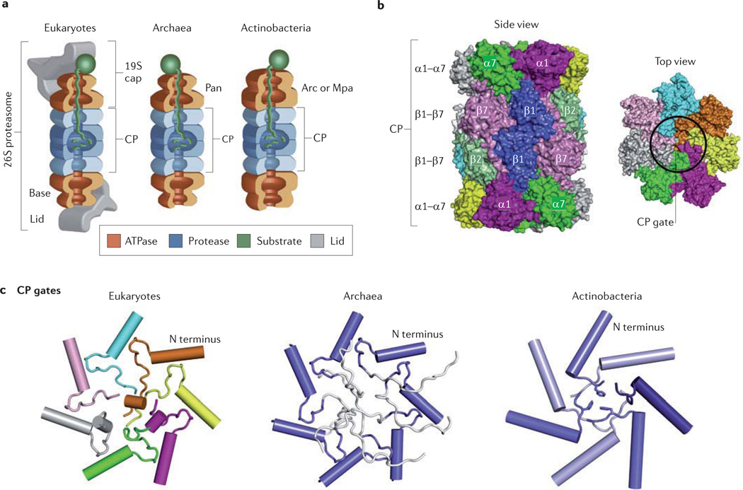
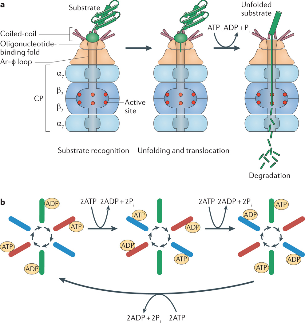
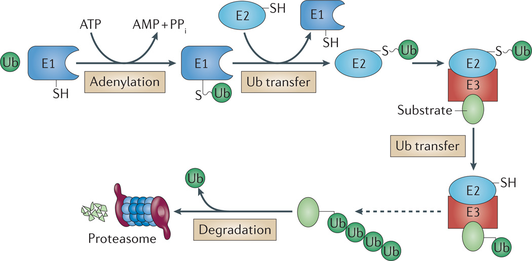
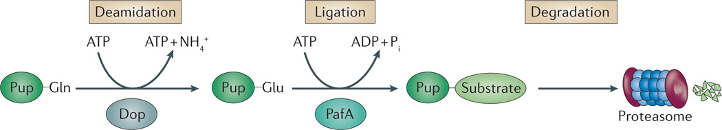
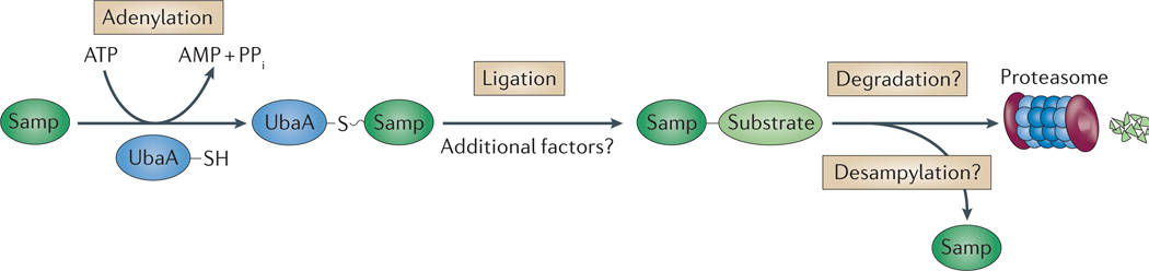
Like other energy-dependent proteases, proteasomes, which are found across the three domains of life, are self-compartmentalized and important in the early steps of proteolysis. Proteasomes degrade improperly synthesized, damaged or misfolded proteins and hydrolyse regulatory proteins that must be specifically removed or cleaved for cell signalling. In eukaryotes, proteins are typically targeted for proteasome-mediated destruction through polyubiquitylation, although ubiquitin-independent pathways also exist. Interestingly, actinobacteria and archaea also covalently attach small proteins (prokaryotic ubiquitin-like protein (Pup) and small archaeal modifier proteins (Samps), respectively) to certain proteins, and this may serve to target the modified proteins for degradation by proteasomes.


Figures

Figure 1
Figure 1. Basic structures of proteasomes across domains of life
a | All proteasomes are composed of a 20S catalytic core particle (CP) formed from four stacked heptameric rings of α- and β-subunits. The CPs can associate with AAA+ ATPases, which unfold and translocate substrate proteins into the CP by an ATP-dependent mechanism. In eukaryotes (using yeast as an example), six different ATPase subunits (Rpt1–Rpt6) form the hexameric ring of the 19S cap, which associates with CPs to form 26S proteasomes. The 19S cap can be separated into base and lid subcomplexes, with the base harbouring the Rpt1–Rpt6 subunits (which use ATP to fuel CP-mediated degradation of folded proteins), and the lid including the deubiquitylating enzyme Rpn11. The proteasomal AAA+ proteins of archaea (proteasome-activating nucleotidase (Pan)) and of actinobacteria (AAA+ ATPase forming a ring-shaped complex (Arc) or mycobacterial proteasome ATPase (Mpa)) assemble into homohexameric rings and associate with CPs in vitro, but the evidence that these ATPases interact with their cognate CPs in vivo is limited. b | Side and top views of a yeast CP provide a perspective on the basic CP structure. c | Across the domains of life, the α-subunit amino-terminal tails that gate the openings of CPs differ in the extent to which they seal the CP channel from substrate entry. Eukaryotic CPs are gated primarily by the well-ordered N-terminal tails of α2, α3 and α4 subunits, which form numerous hydrogen bonds and van der Waals contacts. Gates of archaeal CPs (Thermoplasma acidophlium CPs synthesized in recombinant Escherichia coli) are disordered (residues in white are highly mobile). In actinobacterial CPs, the seven α-subunits are identical but can adopt three different conformations at their N termini (indicated by different shading) to form an ordered closed gate. Part a is modified, with permission, from REF. © (2009) Elsevier. Parts b and c are reproduced, with permission, from REF. © (2011) Elsevier.
Figure 2
Figure 2. Ordered reaction cycle in protein degradation by proteasomes
a | Model of proteolysis based on archaeal proteasome-activating nucleotidase (Pan) and core particle (CP) complexes. The amino-terminal coiled-coil domain of each Pan subunit forms a pair with one of its neighbours. The three tentacle-like coiled-coil pairs protrude from the ATPase face most distal to the CP and surround a pore formed by an oligonucleotide-binding fold, which may serve as an entry point for substrate proteins to traverse into the ATPase channel. An aromatic–hydrophobic (Ar–φ) loop within the narrowest region of the ATPase channel may grip and pull down on substrate proteins, a process driven by ATP hydrolysis. Protein unfolding is thought to occur from these repetitive power strokes, with the oligonucleotide-binding fold providing a rigid platform and narrow opening to stimulate this unfolding. Unfolded protein substrates are translocated through the ATPase channel to the CP for degradation. b | Proteasomal ATPases seem to function as para-subunit pairs in ATP binding, ATP hydrolysis and ADP release during protein unfolding and docking to the CP. ATP binding to a para-subunit pair (red) induces conformational changes in adjacent subunit pairs, so that the clockwise pair (blue) becomes nucleotide free and the anticlockwise pair (green) becomes bound to ADP. Following ATP hydrolysis, the ATP-bound partners (red) are converted to an ADP-bound state, thus simulating the clockwise pair to bind ATP and the anticlockwise pair to release ADP. Thus, an ordered reaction cycle is perpetuated with coordinated conformational changes in para-subunit pairs, probably providing the power strokes for pulling and unfolding the substrates. At any given time, only a subset of the carboxy-terminal HbYX motifs in the ATPase (those in para-subunits bound to ATP) may be extended to open the CP gates. Pi, inorganic phosphate. Part b is modified, with permission, from REF. © (2011) Elsevier.
Figure 3
Figure 3. Ubiquitylation as a signal for degradation
Ubiquitylation is a common signal for eukaryotic 26S proteasomes and involves a cascade of E1 ubiquitin-activating, E2 ubiquitin-conjugating and E3 ubiquitin ligase enzymes. In this cascade, E1 (plus ATP) first adenylates the carboxy-terminal carboxylate of ubiquitin (Ub), forming Ub–AMP, and then forms a Ub thioester intermediate (E1–Ub). Ubiquitin is transferred from E1 to E2, and then to the protein target with assistance from E3 (although ubiquitylation without E3 can occur). Typically, an isopeptide bond is formed between the ubiquitin C-terminal carboxylate and the ε-amino group of a Lys side chain of the substrate protein or the growing ubiquitin chain (Lys48-linked ubiquitin chains are common signals for 26S proteasomes). Deubiquitylating enzymes within 26S proteasomes release and recycle ubiquitin during substrate protein degradation. PPi, inorganic pyrophosphate.
Figure 4
Figure 4. Pupylation as a signal recognized by proteasomes in bacteria
Pupylation and proteasome-mediated proteolysis in actinobacteria. In pupylation, the carboxy-terminal Gln of prokaryotic ubiquitin-like protein (Pup) is deamidated to Glu by Dop. PafA can then attach Pup to substrates, mediating their proteasomal degradation. Once conjugated to protein substrates, Pup binds to the coiled-coil domain of the proteasomal ATPase (called mycobacterial proteasome ATPase (Mpa) in mycobacteria), and a region of Pup is converted from a disordered state into an α-helix (not shown). Pi, inorganic phosphate.
Figure 5
Figure 5. Sampylation and proteasomes in archaea
Similar to ubiquitylation, evidence suggests that the small archaeal modifier proteins (Samps) are adenylated at their carboxy-terminal carboxylate by an E1 ubiquitin-activating-like enzyme (UbaA) and transferred to Lys side chains of protein substrates. Whether additional factors (other than the E1) are needed to ensure proper selection of protein targets and whether sampylated proteins are degraded by proteasomes remain to be determined. Although Lys58-linked Samp2 chains have been detected, it is unclear whether these chains are anchored to substrate proteins (not shown). PPi, inorganic pyrophosphate.

References

    1. Volker C, Lupas AN. Molecular evolution of proteasomes. Curr. Top. Microbiol. Immunol. 2002;268:1–22. - PubMed
    1. Mehnert M, Sommer T, Jarosch E. ERAD ubiquitin ligases: multifunctional tools for protein quality control and waste disposal in the endoplasmic reticulum. Bioessays. 2010;32:905–913. - PubMed
    1. Bengtson MH, Joazeiro CA. Role of a ribosome-associated E3 ubiquitin ligase in protein quality control. Nature. 2010;467:470–473. - PMC - PubMed
    1. Barford D. Structure, function and mechanism of the anaphase promoting complex (APC/C) Q. Rev. Biophys. 2011;44:153–190. - PubMed
    1. Bader M, Steller H. Regulation of cell death by the ubiquitin-proteasome system. Curr. Opin. Cell Biol. 2009;21:878–884. - PMC - PubMed

Publication types