

doi: 10.1007/978-1-4614-0631-0_102.
Identification of pigment epithelium-derived factor receptor (PEDF-R) antibody epitopes
Affiliations
- PMID: 22183409
- PMCID: PMC3901639
- DOI: 10.1007/978-1-4614-0631-0_102
Item in Clipboard
Identification of pigment epithelium-derived factor receptor (PEDF-R) antibody epitopes
Adv Exp Med Biol.
2012.
No abstract available
Figures
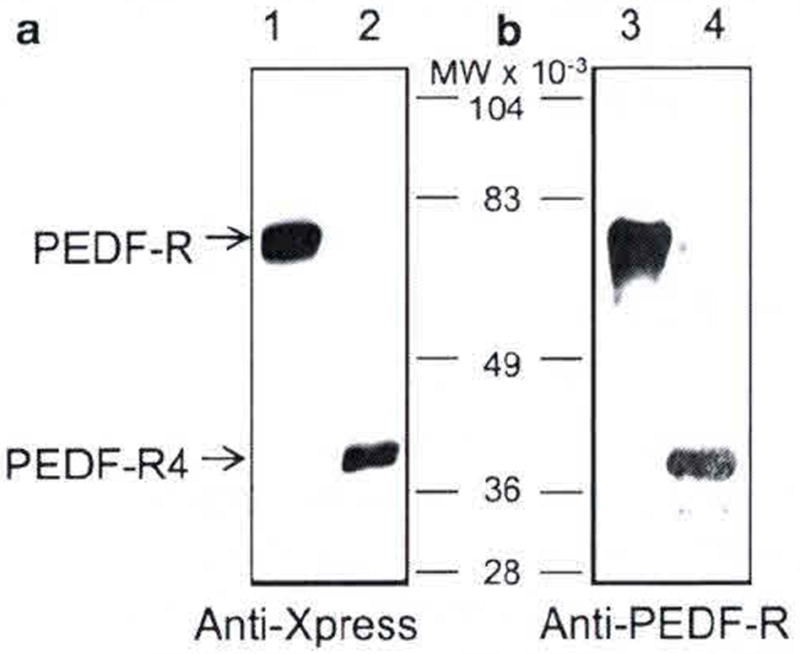
Western blot of recombinant PEDF-R polypeptides. Full-length PEDF-R and C-terminal truncated PEDF-R4 were expressed using in vitro cell-free Escherichia coli expression system. Purified proteins were resolved by SDS-PAGE and electrotransfered to a membrane for immunostaining. Photographs of blots immunostained with anti-Xpress (a) and anti-PEDF-R (b) are shown. Lanes 1 and 3 were PEDF-R, lanes 2 and 4 were PEDF-R4. Migration positions of PEDF-R and PEDF-R4 are indicated with arrows, and of molecular weight markers are in between the two blots
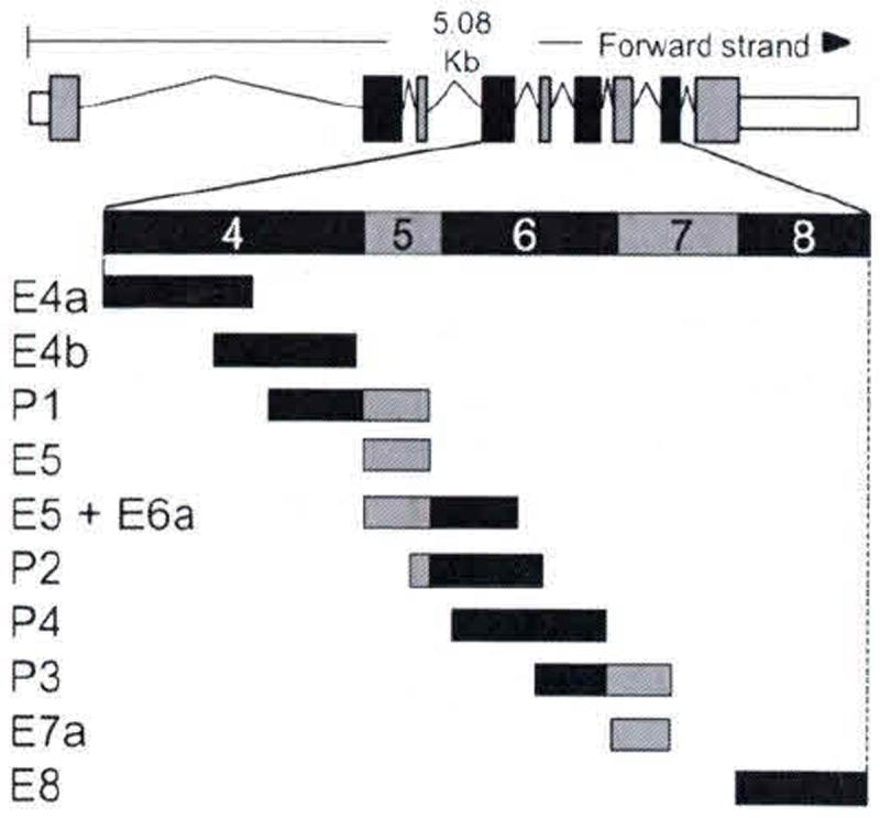
Schematic of rat PEDF-R transcript. Transcript summary information for rat PEDF-R was obtained from http://www.ensembl.org for ENSRNOT00000025319. Exons are illustrated by boxes; coding regions are black and gray; introns are the lines flanking the boxes. Expanded region illustrates the design of synthetic peptides
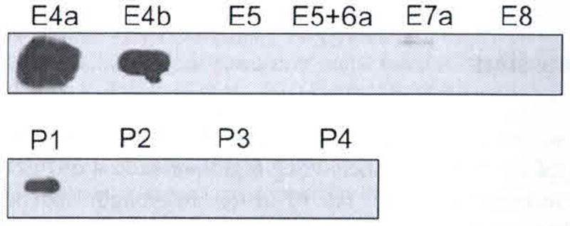
Slot blot of PEDF-R peptides. Peptides (1 mg) were applied to a nitrocellulose membrane using a slot-blot technique and immunostained with anti-PEDF-R. Peptides are indicated to the top of the photograph
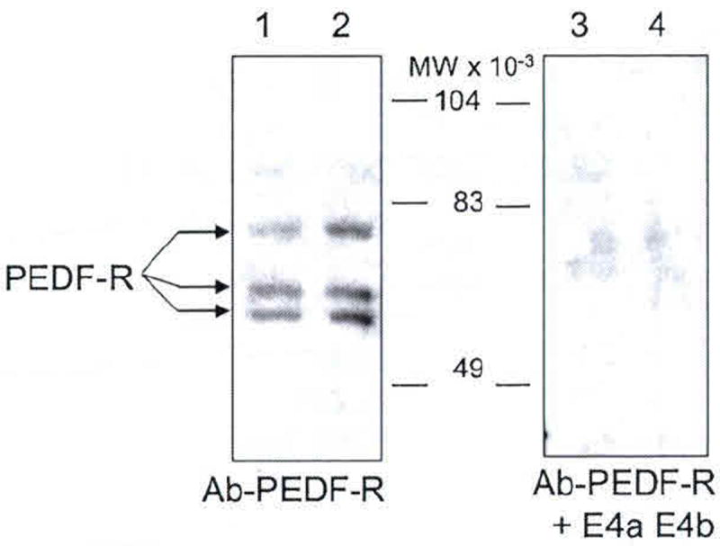
Western blot of native PEDF-R from retina R28 cells. Membrane fractions obtained from R28 cells were resolved by SDS-PAGE. Total protein loaded in lanes 1–4 was 6 μg each. Lanes 1 and 2, and lanes 3 and 4 were replicates. Immunoreactions with anti-PEDF-R were for lanes 1 and 2, and with anti-PEDF-R preincubated with peptides E4a and E4b were for lanes 3 and 4. Migration positions of PEDF-R isoforms are indicated with arrows, and molecular weight markers are in the center
Similar articles
-
Nicotine increases the VEGF/PEDF ratio in retinal pigment epithelium: a possible mechanism for CNV in passive smokers with AMD.Invest Ophthalmol Vis Sci. 2011 Jun 1;52(6):3842-53. doi: 10.1167/iovs.10-6254. Invest Ophthalmol Vis Sci. 2011. PMID: 21330654 Free PMC article.
-
Identification of a lipase-linked cell membrane receptor for pigment epithelium-derived factor.J Biol Chem. 2006 Dec 8;281(49):38022-37. doi: 10.1074/jbc.M600353200. Epub 2006 Oct 10. J Biol Chem. 2006. PMID: 17032652
-
Pigment epithelium-derived factor (PEDF) prevents retinal cell death via PEDF Receptor (PEDF-R): identification of a functional ligand binding site.J Biol Chem. 2013 Aug 16;288(33):23928-42. doi: 10.1074/jbc.M113.487884. Epub 2013 Jul 1. J Biol Chem. 2013. PMID: 23818523 Free PMC article.
-
Delivery Systems of Retinoprotective Proteins in the Retina.Int J Mol Sci. 2021 May 19;22(10):5344. doi: 10.3390/ijms22105344. Int J Mol Sci. 2021. PMID: 34069505 Free PMC article. Review.
-
The applied biochemistry of PEDF and implications for tissue homeostasis.Growth Factors. 2010 Aug;28(4):280-5. doi: 10.3109/08977191003604513. Growth Factors. 2010. PMID: 20166889 Free PMC article. Review.
Cited by
-
PEDF and its roles in physiological and pathological conditions: implication in diabetic and hypoxia-induced angiogenic diseases.Clin Sci (Lond). 2015 Jun;128(11):805-23. doi: 10.1042/CS20130463. Clin Sci (Lond). 2015. PMID: 25881671 Free PMC article. Review.
-
Pigment epithelium-derived factor reduces apoptosis and pro-inflammatory cytokine gene expression in a murine model of focal retinal degeneration.ASN Neuro. 2013 Nov 26;5(5):e00126. doi: 10.1042/AN20130028. ASN Neuro. 2013. PMID: 24160756 Free PMC article.
-
Pigment Epithelium-Derived Factor Promotes Axon Regeneration and Functional Recovery After Spinal Cord Injury.Mol Neurobiol. 2019 Nov;56(11):7490-7507. doi: 10.1007/s12035-019-1614-2. Epub 2019 May 2. Mol Neurobiol. 2019. PMID: 31049830 Free PMC article.
References
-
- Barnstable CJ, Tombran-Tink J. Neuroprotective and antiangiogenic actions of PEDF in the eye: molecular targets and therapeutic potential. Prog Retin Eye Res. 2004;23:561–577. - PubMed
-
- Bazan NG, Marcheselli VL, Hu J, et al. Pigment epithelium-derived growth factor (PEDF) selectively up-regulates NPD1 synthesis and release through the apical side of human RPE cells in primary cultures. Invest Ophthalmol Vis Sci. 2005;46:167.
-
- Bouck N. PEDF: anti-angiogenic guardian of ocular function. Trends Mol Med. 2002;8:330–334. - PubMed
-
- Crawford SE, Stellmach V, Ranalli M, et al. Pigment epithelium-derived factor (PEDF) in neuroblastoma: a multifunctional mediator of Schwann cell antitumor activity. J Cell Sci. 2001;114:4421–4428. - PubMed
MeSH terms
Substances
Grants and funding
LinkOut - more resources
Full Text Sources
Miscellaneous
