

Do patient-specific guides improve coronal alignment in total knee arthroplasty?
- PMID: 22183477
- PMCID: PMC3270188
- DOI: 10.1007/s11999-011-2222-2
Do patient-specific guides improve coronal alignment in total knee arthroplasty?
Erratum in
- Clin Orthop Relat Res. 2012 Apr;470(4):1242
Abstract
Background: Coronal alignment may impact clinical outcome and survivorship in TKA. Patient-specific instrumentation has been developed to restore mechanical or kinematic axis and potentially reduce component malpositioning. Although it is clear these instruments add cost, it is unclear whether they improve alignment.
Questions/purposes: We determined whether the mean coronal alignment after TKA performed with conventional versus patient-specific instrumentation better restored the mechanical and kinematic axes and whether there were more outliers with one of the two methods.
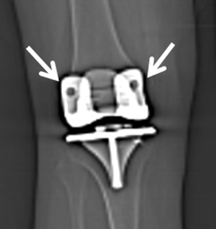
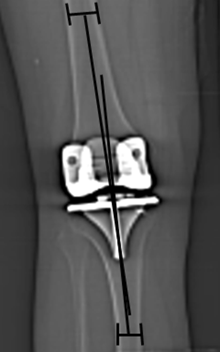
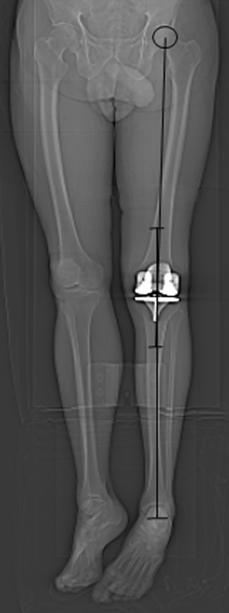
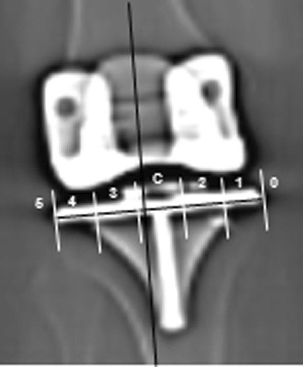
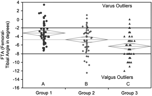
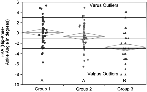
Methods: We retrospectively evaluated 150 primary TKAs performed for osteoarthritis: Group 1 (n = 50) conventional instrumentation; Group 2 (n = 50) patient-specific instrumentation restoring the mechanical axis; Group 3 (n = 50) patient-specific instrumentation restoring the kinematic axis, and measured femorotibial angle, hip-knee-ankle angle, and the zone of the mechanical axis from scout CT images taken 0 to 6 weeks postoperatively.
Results: The mean femorotibial angle differed between the groups: Group 1 had the greatest varus mean alignment and most varus outliers. The mean hip-knee angle was similar between Groups 1 and 2, with Group 3 having greater valgus mean alignment and the most valgus outliers. For the zone of the mechanical axis, Groups 1 and 2 had similar percentages of outliers (40% versus 32%), whereas Group 3 had a greater number of outliers (64%) that were valgus.
Conclusions: TKAs with patient-specific instrumentation restoring the mechanical axis had a similar number of outliers as conventional instrumentation with both groups having more varus outliers than TKAs with patient-specific instrumentation restoring kinematic axis, which had more valgus outliers. Therefore, additional studies are needed to determine whether patient-specific instrumentation improves clinical function or patient satisfaction and whether their routine use can be justified in primary TKA.
Level of evidence: Level III, therapeutic study. See Guidelines for Authors for a complete description of levels of evidence.
Figures

References
-
- Bargren JH, Blaha JD, Freeman MA. Alignment in total knee arthroplasty. Correlated biomechanical and clinical observations. Clin Orthop Relat Res. 1983;173:178–183. - PubMed
-
- Bolognesi MP, Pearle AD, Oryhon JM, Nunley RM. New technology for knee arthroplasty. In: Glassman AH, Lachiewicz PF, Tanzer M, editors. Orthopaedic Knowledge Update: Hip and Knee Reconstruction 4. Rosemont, IL: American Academy of Orthopaedic Surgeons; 2011. pp. 119–137.
Publication types
MeSH terms
LinkOut - more resources
Full Text Sources
Medical
Research Materials
