. 2012 Feb 15;590(4):809-25.
doi: 10.1113/jphysiol.2011.222836. Epub 2011 Dec 19.

Ca(v)2 channels mediate low and high voltage-activated calcium currents in Drosophila motoneurons

Affiliations

Ca(v)2 channels mediate low and high voltage-activated calcium currents in Drosophila motoneurons

Stefanie Ryglewski et al. J Physiol. .

Abstract

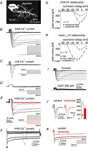
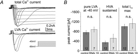
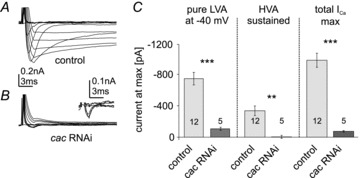
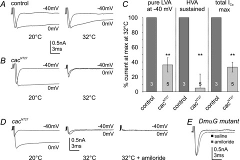
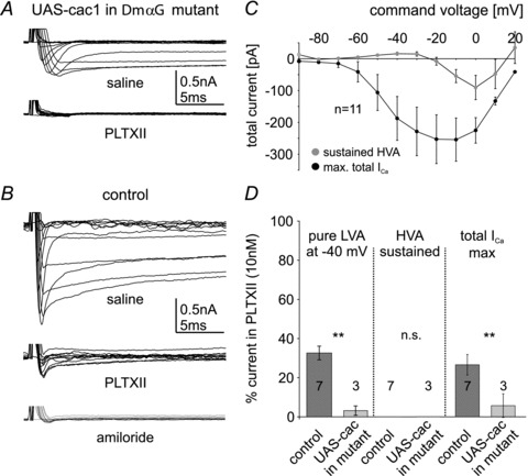
Different blends of membrane currents underlie distinct functions of neurons in the brain. A major step towards understanding neuronal function, therefore, is to identify the genes that encode different ionic currents. This study combined in situ patch clamp recordings of somatodendritic calcium currents in an identified adult Drosophila motoneuron with targeted genetic manipulation. Voltage clamp recordings revealed transient low voltage-activated (LVA) currents with activation between –60 mV and –70 mV as well as high voltage-activated (HVA) current with an activation voltage around –30 mV. LVA could be fully inactivated by prepulses to –50 mV and was partially amiloride sensitive. Recordings from newly generated mutant flies demonstrated that DmαG (Ca(v)3 homolog) encoded the amiloride-sensitive portion of the transient LVA calcium current. We further demonstrated that the Ca(v)2 homolog, Dmca1A, mediated the amiloride-insensitive component of LVA current. This novel role of Ca(v)2 channels was substantiated by patch clamp recordings from conditional mutants, RNAi knock-downs, and following Dmca1A overexpression. In addition, we show that Dmca1A underlies the HVA somatodendritic calcium currents in vivo. Therefore, the Drosophila Ca(v)2 homolog, Dmca1A, underlies HVA and LVA somatodendritic calcium currents in the same neuron. Interestingly, DmαG is required for regulating LVA and HVA derived from Dmca1A in vivo. In summary, each vertebrate gene family for voltage-gated calcium channels is represented by a single gene in Drosophila, namely Dmca1D (Ca(v)1), Dmca1A (Ca(v)2) and DmαG (Ca(v)3), but the commonly held view that LVA calcium currents are usually mediated by Ca(v)3 rather than Ca(v)2 channels may require reconsideration.


Figures

Figure 1
Figure 1. Generation and validation of DmαG excision mutants
A, illustration of the Ca-alpha1T gene (DmαG) showing the placement of inserted piggyBac elements. Placement of piggyBac elements on Ca-alpha1T gene span are indicated by blue arrows. Transcript variant B is shown as an example transcript indicating the placement of the exons derived from Ca-alpha1T, seen here as purple boxes. The yellow box indicates the region of the gene that is expected to be absent after the deletion has occurred. B, confirmation of deletion on the X chromosome of a Ca-alpha 1T mutant male individual. Columns A and B show results of two-sided PCR confirming the presence of both piggyBac elements in the mutant, using transposon-specific and genomic primers designed for PBac{WH}f05809 and PBac{WH}Ca-α1Tf06496, respectively. Columns C and D show the presence of the two elements in the respective stock lines. Columns E–G show the absence of the DNA region between the piggyBac elements in the mutant individual, using primers that were designed for the PBac{WH}f05809 stock line. One primer was located within the region of DNA that was to be deleted and the other primer was located on a portion of the PBac{WH}f05809 element expected to be present after the deletion occurred. Column E shows that the putative mutant does not have a fragment at the expected band size of 1500 bp. Sample F shows that the PBac{WH}f05809 stock line does have a fragment at the expected size. Sample G shows Canton-S wildtype. Note that the residual bands that are seen in sample E are not due to the presence of the PBac{WH}f05809 element. The PCR annealing temperature and extension time for the samples that are in columns A–D were 62°C and 1 min, respectively. The PCR annealing temperature and extension time for the samples in columns E–G were 66°C and 2 min, respectively.
Figure 2
Figure 2. Low voltage-activated (LVA) and high voltage-activated (HVA) calcium current in motoneuron 5
The monopolar motoneuron 5, MN5, stained intracellularly with dextran-tetramethylrhodamine reveals a complex dendritic tree (A) that is comprised of more than 6500 μm of dendrititic length. The axon projects contralaterally towards the dorsolongitudinal flight muscle. Whole-cell recordings were conducted from the soma of MN5. Command voltage steps from –90 mV to +20 mV in 10 mV increments from a holding potential of –90 mV reveal transient LVA and sustained HVA currents (B). LVA calcium current is transient (inactivates within 5 ms after pulse onset), activates between –70 mV and –60 mV (C) and is completely inactivated following a 1 s lasting prepulse to –50 mV (D). HVA is electrically isolated by a prepulse to –50 mV of 1 s duration followed by command voltage steps from –90 mV to +20 mV in 10 mV increments (E and F). HVA activates between –40 mV and –30 mV within 5 ms after pulse onset (E) and does not fully inactivate during the 200 ms lasting voltage step (F). Command voltages at which HVA does not yet activate and LVA is fully inactivated following a 1 s lasting prepulse to –50 mV are shown in red (E). Current–voltage plot in G depicts activation of isolated HVA at peak shortly after pulse onset (F and G; black circles, maximum HVA) and as mean sustained current between 100 ms and 120 ms after pulse onset (F, G; grey circles, sustained HVA). Maximum mean current amplitude is at –10 mV. Current–voltage plot in H shows isolated LVA calcium current between command voltages of –90 mV and –40 mV (H; red circles), maximum total calcium current (max. total ICa, sum of LVA and HVA) between command potentials more depolarized than –40 mV and +20 mV as measured at peak shortly after pulse onset (H; black circles). The sustained portion of HVA as measured between 100 ms and 120 ms after pulse onset is plotted for voltage steps from a holding of –90 mV (H; grey circles, sustained ICa). Cadmium (300 μm) blocks all calcium current as evoked by voltage steps from –90 mV to +20 mV from a holding potential of –90 mV (I). Amiloride (1 mm) blocks a portion of the LVA calcium current as compared with control (J, voltage steps –60, –50 and –40 mV are shown; black, control; red, amiloride). Quantification shows that amiloride reduces LVA peak current amplitude at –40 mV from –756 ± 152 pA in control to –498 ± 127 pA in amiloride (J). Amiloride has no obvious effect on electrically isolated HVA current (K). Data are represented as mean ± SEM. Insets in B–E and K depict voltage steps conducted to evoke calcium currents.
Figure 3
Figure 3. Dmca1D does not contribute to somatodendritic calcium current in MN5
A, whole-cell total calcium current in Dmca1D RNAi knock-down animals as evoked by voltage steps from –90 mV to +20 mV from a holding potential of –90 mV in 10 mV increments (top trace) and isolated LVA current at command potentials from –90 mV to –40 mV (bottom trace). Calcium currents are not affected by Dmca1D RNAi knock-down as compared with control (compare with Fig. 2B and C). B, quantitative comparison of isolated LVA calcium current amplitudes at –40 mV (left column), sustained HVA (middle column), and maximum total calcium current amplitudes (total ICa max, right column) in controls (dark grey bars) and following Dmca1D RNAi knock-down (light grey bars) reveals no effect of Dmca1D RNAi knock-down (n.s., unpaired Student's t test). Data are shown as mean ± SEM.
Figure 4
Figure 4. Multiple calcium current phenotypes occur in DmαG null mutants
A, 56% of all DmαG mutants recorded exhibit no measurable calcium current (A, first bar in D); B, 28% show both LVA and HVA calcium current with reduced amplidues (B, second bar in D); and C, 16% exhibit control-like currents (C, third bar in D). E, on average, isolated LVA calcium current at –40 mV (left column), sustained HVA (middle column), and total calcium current at maximum (total ICa max, right column) are significantly reduced in DmαG excision mutants as compared with control (Mann–Whitney U test, ***P < 0.001; **P < 0.01). Data are shown as median with 25% and 75% quartiles. Dotted error bars depict maximum and minimum values.
Figure 5
Figure 5. Dmca1A RNAi knock-down reduces LVA and eliminates HVA currents
Representative recordings of MN5 calcium currents in control (A) and following targeted expression of UAS-Dmca1A RNAi; UAS-dicer (B). Remaining small outward current after pulse onset at more depolarized command voltages probably reflects potassium current that was un-masked by knock-down of inward current. Inset in B shows enlargement of current in UAS-Dmca1A RNAi for selected command voltages between –90 mV and –40 mV to show the LVA current that remained after Dmca1A RNAi knock-down. Quantification (C) shows that both isolated LVA current at –40 mV (left column) and maximum total calcium current (total ICa max, right column) are reduced by more than 80% (unpaired Student's t test, ***P < 0.001) as compared with control, and sustained HVA current is eliminated following Dmca1A knock-down (**P < 0.01). Light grey bars represent control, dark grey bars represent Dmca1A RNAi knock-down. Data represent mean ± SEM.
Figure 6
Figure 6. Conditional Dmca1A knock-down in the adult MN5 reduces LVA and abolishes HVA calcium currents
AC, calcium currents as evoked by voltage steps to –40 mV and 0 mV before and after bath temperature shifts (20°C and 32°C) in control (A) and in conditional mutant cacNT27 animals (B). Isolated LVA at –40 mV (left column), sustained HVA (middle column), and maximum total calcium current amplitudes (right column) are not significantly affected by temperature shifts in controls (C, dark grey bars). In cacNT27 animals isolated LVA calcium current at –40 mV, sustained HVA and maximum total calcium current amplitudes are similar to controls at 20°C, but at 32°C isolated LVA at –40 mV (left column) and total calcium current amplitudes (right column) are significantly reduced (by 63.69 ± 9%, and 64.23 ± 6%, respectively; Student's t test, **P < 0.01, light grey bars). HVA calcium current is virtually abolished at 32°C (C, middle column, **P < 0.01, light grey bar). Application of amiloride (1 mm) at 32°C in cacNT27 mutants abolishes the small LVA calcium current remaining after temperature shift of cacNT27 mutants to 32°C (D). Amiloride does not affect the isolated LVA calcium current amplitude at –40 mV in DmαG mutants (E). Data are shown as mean ± SEM.
Figure 7
Figure 7. Cacophony can produce LVA and HVA calcium current in vivo in the absence of DmαG
Expression of UAS-cac1 in MN5 in a DmαG mutant background yielded LVA and HVA calcium currents (A, top trace) with normal activation voltages (C) but reduced amplitudes as compared with controls (compare C and Fig. 2H). In this genotype both isolated LVA at –40 mV (D, left column, light grey bar) as well as total maximum calcium current (A, right column in D, light grey bar) were strongly reduced and HVA was abolished by bath application of PLTXII (bottom trace in A, middle column in D, light grey bar). In controls PLTXII blocks a large portion of LVA (left column in D, dark grey bar) and abolishes all HVA calcium current (middle trace in B, middle column in D). The LVA calcium current remaining after PLTXII is blocked by amiloride (1 mm, bottom trace in B). In DmαG mutants with cac1 expression in MN5 significantly more isolated LVA at –40 mV (left column in D, light grey bar) as well as total maximum calcium current (right column in D, light grey bar) are blocked by PLTXII than in control (D, left and right columns, dark grey bars; Student's t test, **P < 0.01). Data are shown as mean ± SEM.

Similar articles

Cited by

References

    1. Baines RA, Bate M. Electrophysiological development of central neurons in the Drosophila embryo. J Neurosci. 1998;18:4673–4683. - PMC - PubMed
    1. Baines RA, Pym EC. Determinants of electrical properties in developing neurons. Semin Cell Dev Biol. 2006;17:12–19. - PubMed
    1. Bleakman D, Bowman D, Bath CP, Brust PF, Johnson EC, Deal CR, Miller RJ, Ellis SB, Harpold MM, Hans M, Grantham CJ. Characteristics of a human N-type calcium channel expressed in HEK293 cells. Neuropharmacology. 1995;34:753–765. - PubMed
    1. Bock G, Gebhart M, Scharinger A, Jangsangthong W, Busquet P, Poggiani C, S Sartori, Mangoni ME, Sinnegger-Brauns MJ, Herzig S, Striessnig J, Koschak A. Functional properties of a newly identified C-terminal splice variant of Cav1.3 L-type Ca2+ channels. J Biol Chem. 2011;286:42736–42748. - PMC - PubMed
    1. Boerner J, Duch C. Average shape standard atlas for the adult Drosophila ventral nerve cord. J Comp Neurol. 2010;518:2437–2455. - PubMed

Publication types

LinkOut - more resources