Review
. 2012 May 9;18(1):486-96.
doi: 10.2119/molmed.2011.00414.

The receptor that tames the innate immune response

Affiliations
Review

The receptor that tames the innate immune response

Michael Brines et al. Mol Med. .

Abstract

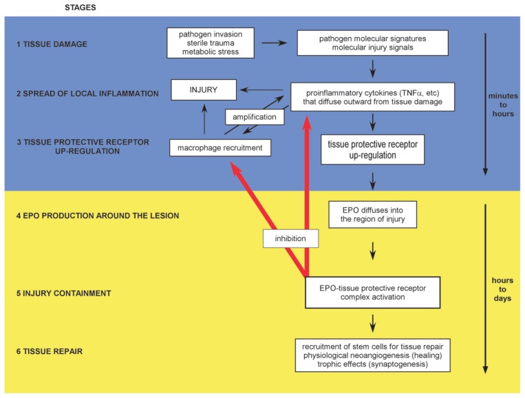
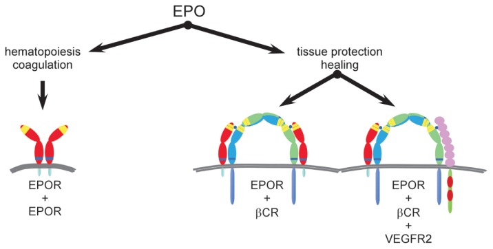
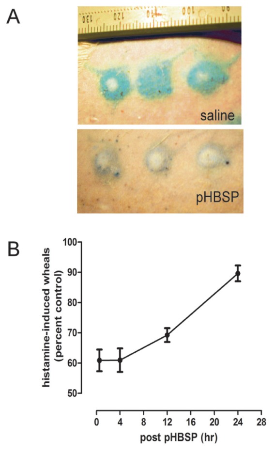
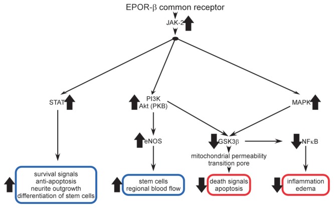
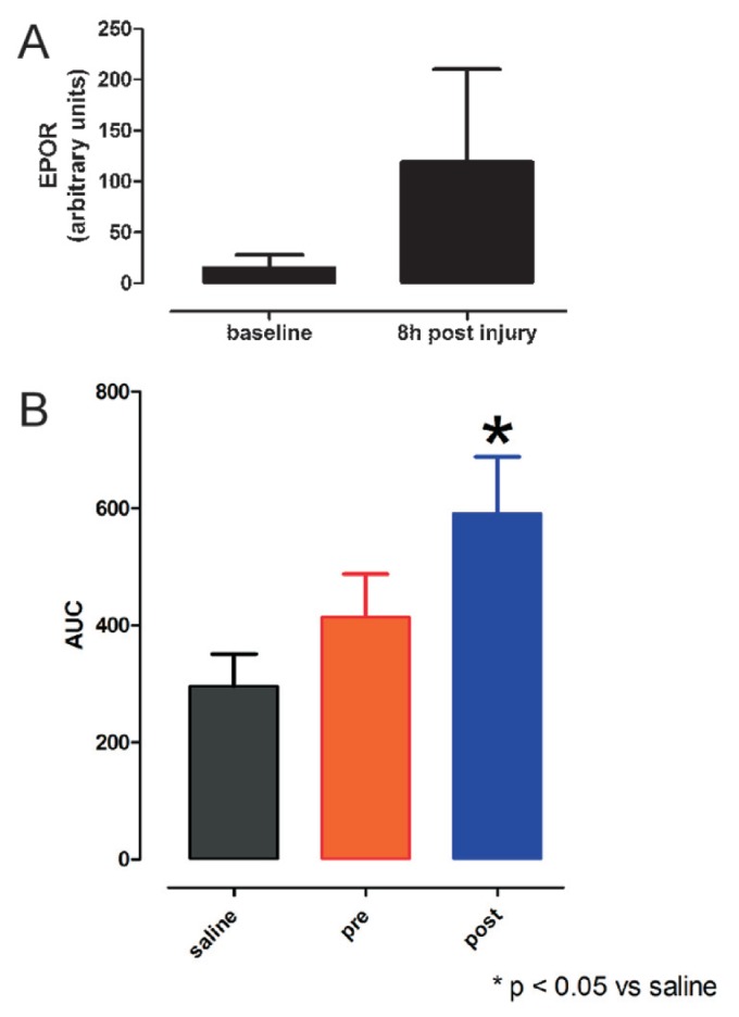
Tissue injury, hypoxia and significant metabolic stress activate innate immune responses driven by tumor necrosis factor (TNF)-α and other proinflammatory cytokines that typically increase damage surrounding a lesion. In a compensatory protective response, erythropoietin (EPO) is synthesized in surrounding tissues, which subsequently triggers antiinflammatory and antiapoptotic processes that delimit injury and promote repair. What we refer to as the sequelae of injury or disease are often the consequences of this intentionally discoordinated, primitive system that uses a "scorched earth" strategy to rid the invader at the expense of a serious lesion. The EPO-mediated tissue-protective system depends on receptor expression that is upregulated by inflammation and hypoxia in a distinctive temporal and spatial pattern. The tissue-protective receptor (TPR) is generally not expressed by normal tissues but becomes functional immediately after injury. In contrast to robust and early receptor expression within the immediate injury site, EPO production is delayed, transient and relatively weak. The functional EPO receptor that attenuates tissue injury is distinct from the hematopoietic receptor responsible for erythropoiesis. On the basis of current evidence, the TPR is composed of the β common receptor subunit (CD131) in combination with the same EPO receptor subunit that is involved in erythropoiesis. Additional receptors, including that for the vascular endothelial growth factor, also appear to be a component of the TPR in some tissues, for example, the endothelium. The discoordination of the EPO response system and its relative weakness provide a window of opportunity to intervene with the exogenous ligand. Recently, molecules were designed that preferentially activate only the TPR and thus avoid the potential adverse consequences of activating the hematopoietic receptor. On administration, these agents successfully substitute for a relative deficiency of EPO production in damaged tissues in multiple animal models of disease and may pave the way to effective treatment of a wide variety of insults that cause tissue injury, leading to profoundly expanded lesions and attendant, irreversible sequelae.


Figures

Figure 1
Figure 1
The innate tissue-protective response is characterized by distinct stages in which pro- and antiinflammatory cytokines and their receptors are temporally and spatially distinct. Stage 1: Pathogen-associated molecular patterns, hypoxia and molecular injury signals trigger the innate immune response driven by proinflammatory cytokines. Stage 2: Proinflammatory cytokines are produced within the injury region and diffuse into the surrounding normal tissue, priming it for self-destruction (the penumbra). Stage 3: Simultaneously, upregulation of the TPR by proinflammatory cytokines occurs within the penumbra. Macrophages recruited into the damaged area amplify injury, and the lesion progressively enlarges as the inflammatory components self-amplify and diffuse outward, causing additional damage. The high levels of proinflammatory cytokines suppress production of EPO, the ligand of the TPR. Stage 4: EPO is produced at the lesion periphery, where proinflammatory cytokine concentrations are lower, and diffuses inward, engaging the TPR, which in turn inhibits proinflammatory cytokine production and rescues cells within the penumbra from apoptosis. Stage 5: The lesion size is contained at the boundary defined by the effective inhibition of apoptosis and unrescuable cellular destruction. Stage 6: In the subacute phase, tissue receptor activation also mobilizes tissue-specific and endothelial stem cells that participate in angiogenesis and other aspects of repair. Stages 1–3 occur rapidly (minutes to hours), while stages 4–6 occur with a substantial time delay (hours to days).
Figure 2
Figure 2
Administration of EPO rescues the penumbra after cerebral ischemia. An ischemic core (IC; panel E) is caused by a 1-h occlusion of the middle cerebral artery distal to the rhinal artery in a rat, followed by reperfusion (stage 1 injury response, performed according to the methodology seen in references and 81). TNFα and other proinflammatory cytokines are produced within this central volume of injury corresponding to the circulatory territory of the middle cerebral artery (MCA) and diffuse into the adjacent cerebral cortex to the right of IC (stage 2 injury response). Microglia (the macrophage equivalents of the nervous system) are recruited into the penumbra (right panels) and amplify injury. Within the penumbra, the TPR is subsequently upregulated (stage 3 injury response), but upregulation of its ligand EPO is suppressed. Therefore, EPO does not penetrate very far into the penumbra from the unsuppressed periphery, producing a large lesion when evaluated 24 h later (light gray area, panel S). However, parenteral administration of EPO (panel E) rescues the penumbra by delivering TPR ligand into this region primed for TPR activity but deficient in endogenous EPO.
Figure 3
Figure 3
EPO uses receptor isoforms for signaling. A homodimer receptor (EPOR)2 is formed by the spontaneous self-assembly of EPOR monomers and mediates erythropoiesis (left). The receptor-ligand stoichiometry is 2:1 for EPOR and EPO, respectively. The hematopoietic receptor is characterized by a high affinity for EPO and requires constant, low circulating concentrations of EPO to maintain adequate erythrocyte production. Other biological activities controlled by (EPOR)2 are those associated with preservation of red cell mass in the setting of hemorrhage. These functions include induction of a prothrombotic state by activation of vascular endothelium adhesion molecules and accelerated maturation of megakaryocytes, as well as stimulation of vascular smooth muscle leading to shunting of blood from noncritical tissues. All of these activities serve to minimize blood loss. In contrast, available evidence supports the existence of a different receptor for tissue protection (middle) that involves assembly of EPOR and βCR subunits, as well as other receptor subunits (right; for example, VEGFR2) in certain tissues. On the basis of analogy to the GM-CSF receptor, the stoichiometry may be in a 2:2:2 ratio for EPO, EPOR and βCR. The TPR is characterized by a lower affinity for the endogenous ligand EPO and requires only brief receptor occupancy to initiate long-lasting biological effects. Because high doses of EPO are required for TPR activation, tissue protection by exogenous EPO is unavoidably accompanied by these potential serious adverse consequences. To circumvent these potential problems, analogs of EPO have been engineered that lack hematopoietic potency but retain tissue-protective activity. This strategy provides tissue protection while avoiding the significant potential side effects of using EPO itself.
Figure 4
Figure 4
The TPR is characterized by expression of βCR and EPOR. (A) A variety of neurons within rat spinal cord ventral horn express both βCR and EPOR. (B) In this experiment, wild-type rat cardiomyocytes undergo apoptosis after exposure to stuarosporine in vitro (column WT). Addition of EPO protects from staurosporine (WT+EPO). In contrast, cardiomyocytes obtained from βCR KO (βc KO) animals are not protected by EPO (βc KO+EPO). (****P < 0.001 versus staurosporine alone; reproduced with adaptation from [35]).
Figure 5
Figure 5
The TPR acts a molecular switch activating long-lasting tissue protection. Histamine injected intradermally activates histamine receptors that within several minutes cause intense vasoconstriction at the site of injection (wheal) that is surrounded by extravasated intravascular proteins (flare). (A) This reaction can be quantified by labeling albumin with Evans blue dye. As an example, a rat that received saline 4 h earlier exhibited a vigorous response to histamine (top). In contrast, a rat that received pHBSP 4 h earlier responded with substantially less vascular extravasation, as indicated by the smaller region less intensively labeled with Evans blue (bottom). (B) A single dose of the tissue-protective molecule pHBSP (30 μg/kg) exerts long-lasting effects, antagonizing histamine administered hours later (top; ± SEM). (Please see text for details.)
Figure 6
Figure 6
Multiple signaling pathways have been documented for the TPR. A variety of signaling pathways activated by the TPR have been determined using the nonerythropoietic tissue-protective compounds CEPO and pHBSP. Tissue-protective receptor occupancy leads to JAK-2 phosphorylation which subsequently activates a number of divergent cascades. In some tissues, these pathways overlap and are therefore redundant; but in others, each appears to serve specific functions. Both enhanced (blue boxes) and suppressed (red boxes) responses have been identified (see text for discussion).
Figure 7
Figure 7
Tissue protection depends on upregulation of the TPR. (A) Rat spinal cord does not express high levels of EPOR, as estimated by immunocytochemistry. However, within a few hours after a 1-min compression of the spinal cord at level thoracic 3 (T3), EPOR immunoreactivity more than quadruples (± SEM). (B) AsialoEPO is a tissue-protective molecule derived by removing the terminal sialic acids of the oligosaccharide chains of EPO, which results in a circulating half-life of only ~2 min in vivo. However, tissue-protective activity of asialoEPO is similar to that of EPO. AsialoEPO (50 μg/kg) administered intraperitoneally 24 h before a 1-min compression at T3 in rats is associated with little protection, as assessed by a motor score derived by the area under the curve (AUC) for weekly measurements carried out to 28 d. In contrast, when asialoEPO was administered immediately after release of compression, there was a significant improvement in the motor score compared with control animals (± SEM; drawn from data presented in [87]).

Similar articles

Cited by

References

    1. Kedzierski L, Montgomery J, Curtis J, Handman E. Leucine-rich repeats in host-pathogen interactions. Arch Immunol Ther Exp (Warsz) 2004;52:104–12. - PubMed
    1. Janssens S, Beyaert R. Role of Toll-like receptors in pathogen recognition. Clin Microbiol Rev. 2003;16:637–46. - PMC - PubMed
    1. Andersson U, Tracey KJ. HMGB1 is a therapeutic target for sterile inflammation and infection. Annu Rev Immunol. 2011;29:139–62. - PMC - PubMed
    1. Dzik JM. The ancestry and cumulative evolution of immune reactions. Acta Biochim Pol. 2010;57:443–66. - PubMed
    1. Huffaker A, Pearce G, Ryan CA. An endogenous peptide signal in Arabidopsis activates components of the innate immune response. Proc Natl Acad Sci U S A. 2006;103:10098–103. - PMC - PubMed

LinkOut - more resources