

Targeted chromosomal duplications and inversions in the human genome using zinc finger nucleases
- PMID: 22183967
- PMCID: PMC3290789
- DOI: 10.1101/gr.129635.111
Targeted chromosomal duplications and inversions in the human genome using zinc finger nucleases
Abstract
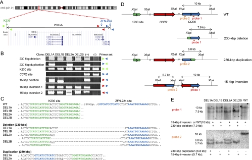
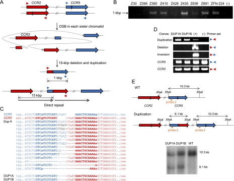
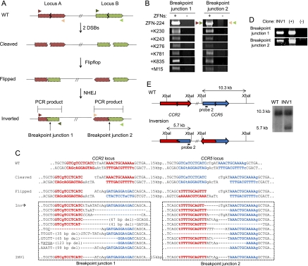
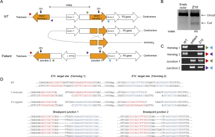
Despite the recent discoveries of and interest in numerous structural variations (SVs)--which include duplications and inversions--in the human and other higher eukaryotic genomes, little is known about the etiology and biology of these SVs, partly due to the lack of molecular tools with which to create individual SVs in cultured cells and model organisms. Here, we present a novel method of inducing duplications and inversions in a targeted manner without pre-manipulation of the genome. We found that zinc finger nucleases (ZFNs) designed to target two different sites in a human chromosome could introduce two concurrent double-strand breaks, whose repair via non-homologous end-joining (NHEJ) gives rise to targeted duplications and inversions of the genomic segments of up to a mega base pair (bp) in length between the two sites. Furthermore, we demonstrated that a ZFN pair could induce the inversion of a 140-kbp chromosomal segment that contains a portion of the blood coagulation factor VIII gene to mimic the inversion genotype that is associated with some cases of severe hemophilia A. This same ZFN pair could be used, in theory, to revert the inverted region to restore genomic integrity in these hemophilia A patients. We propose that ZFNs can be employed as molecular tools to study mechanisms of chromosomal rearrangements and to create SVs in a predetermined manner so as to study their biological roles. In addition, our method raises the possibility of correcting genetic defects caused by chromosomal rearrangements and holds new promise in gene and cell therapy.
Figures
Similar articles
-
Targeted deletion and inversion of tandemly arrayed genes in Arabidopsis thaliana using zinc finger nucleases.G3 (Bethesda). 2013 Oct 3;3(10):1707-15. doi: 10.1534/g3.113.006270. G3 (Bethesda). 2013. PMID: 23979943 Free PMC article.
-
Targeted chromosomal deletions in human cells using zinc finger nucleases.Genome Res. 2010 Jan;20(1):81-9. doi: 10.1101/gr.099747.109. Epub 2009 Dec 1. Genome Res. 2010. PMID: 19952142 Free PMC article.
-
Targeted inversion and reversion of the blood coagulation factor 8 gene in human iPS cells using TALENs.Proc Natl Acad Sci U S A. 2014 Jun 24;111(25):9253-8. doi: 10.1073/pnas.1323941111. Epub 2014 Jun 9. Proc Natl Acad Sci U S A. 2014. PMID: 24927536 Free PMC article.
-
Genome engineering with zinc-finger nucleases.Genetics. 2011 Aug;188(4):773-82. doi: 10.1534/genetics.111.131433. Genetics. 2011. PMID: 21828278 Free PMC article. Review.
-
Advances in genetic modification of farm animals using zinc-finger nucleases (ZFN).Chromosome Res. 2015 Feb;23(1):7-15. doi: 10.1007/s10577-014-9451-7. Chromosome Res. 2015. PMID: 25596823 Review.
Cited by
-
Biological and biomedical applications of engineered nucleases.Mol Biotechnol. 2013 Sep;55(1):54-62. doi: 10.1007/s12033-012-9613-9. Mol Biotechnol. 2013. PMID: 23089945 Review.
-
Cancer translocations in human cells induced by zinc finger and TALE nucleases.Genome Res. 2013 Jul;23(7):1182-93. doi: 10.1101/gr.147314.112. Epub 2013 Apr 8. Genome Res. 2013. PMID: 23568838 Free PMC article.
-
Targeted deletion and inversion of tandemly arrayed genes in Arabidopsis thaliana using zinc finger nucleases.G3 (Bethesda). 2013 Oct 3;3(10):1707-15. doi: 10.1534/g3.113.006270. G3 (Bethesda). 2013. PMID: 23979943 Free PMC article.
-
A mechanistic study on the tolerance of PAM distal end mismatch by SpCas9.J Biol Chem. 2024 Jul;300(7):107439. doi: 10.1016/j.jbc.2024.107439. Epub 2024 Jun 3. J Biol Chem. 2024. PMID: 38838774 Free PMC article.
-
Digenome-seq: genome-wide profiling of CRISPR-Cas9 off-target effects in human cells.Nat Methods. 2015 Mar;12(3):237-43, 1 p following 243. doi: 10.1038/nmeth.3284. Epub 2015 Feb 9. Nat Methods. 2015. PMID: 25664545
References
-
- Bagnall RD, Waseem N, Green PM, Giannelli F 2002. Recurrent inversion breaking intron 1 of the factor VIII gene is a frequent cause of severe hemophilia A. Blood 99: 168–174 - PubMed
-
- Bueno C, Catalina P, Melen GJ, Montes R, Sanchez L, Ligero G, Garcia-Perez JL, Menendez P 2009. Etoposide induces MLL rearrangements and other chromosomal abnormalities in human embryonic stem cells. Carcinogenesis 30: 1628–1637 - PubMed
-
- Bylund L, Kytola S, Lui WO, Larsson C, Weber G 2004. Analysis of the cytogenetic stability of the human embryonal kidney cell line 293 by cytogenetic and STR profiling approaches. Cytogenet Genome Res 106: 28–32 - PubMed
Publication types
MeSH terms
Substances
LinkOut - more resources
Full Text Sources
Other Literature Sources