. 2012 Jan 3;109(1):197-202.
doi: 10.1073/pnas.1104767109. Epub 2011 Dec 19.

Checkpoint kinase 1 (Chk1)-short is a splice variant and endogenous inhibitor of Chk1 that regulates cell cycle and DNA damage checkpoints

Affiliations

Checkpoint kinase 1 (Chk1)-short is a splice variant and endogenous inhibitor of Chk1 that regulates cell cycle and DNA damage checkpoints

Navjotsingh Pabla et al. Proc Natl Acad Sci U S A. .

Abstract

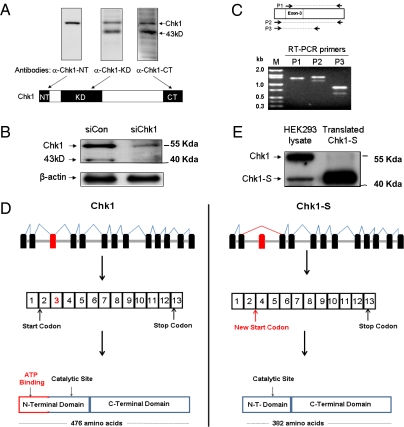
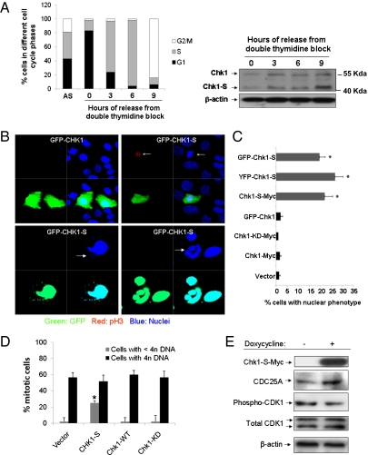
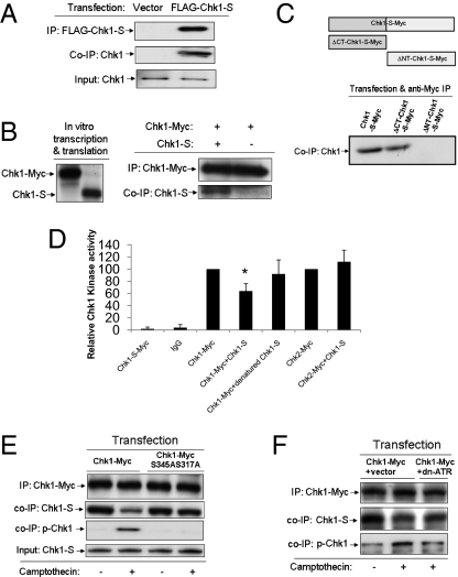
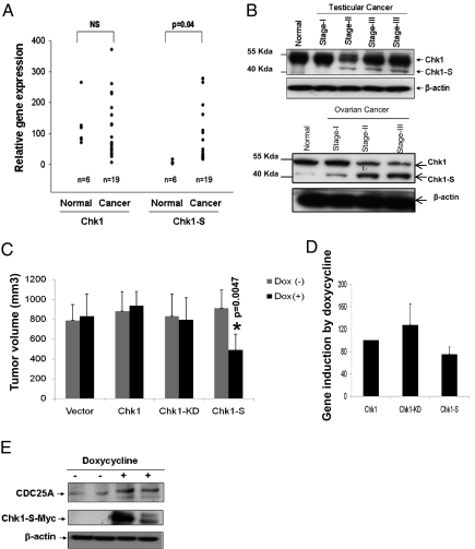
Checkpoint kinase 1 (Chk1) is a key regulator of checkpoint signaling in both the unperturbed cell cycle and DNA damage response. Under these conditions, Chk1 becomes active to prevent premature CDK1 activation and mitotic entry until DNA is properly replicated or repaired. It is unclear how Chk1 activity is controlled in the unperturbed cell cycle. During DNA damage, Chk1 is activated by ataxia telangiectasia and Rad3 related (ATR)-mediated phosphorylation; however, it is not entirely clear how this phosphorylation results in Chk1 activation. Here we report an N-terminally truncated alternative splice variant of Chk1, Chk1-S. Importantly, we show that Chk1-S is an endogenous repressor and regulator of Chk1. In the unperturbed cell cycle, Chk1-S interacts with and antagonizes Chk1 to promote the S-to-G2/M phase transition. During DNA damage, Chk1 is phosphorylated, which disrupts the Chk1-Chk1-S interaction, resulting in free, active Chk1 to arrest the cell cycle and facilitate DNA repair. Higher levels of Chk1-S are expressed, along with Chk1, in fetal and cancer tissues than in normal tissues. However, forced overexpression of Chk1-S in cultured cells and tumor xenografts induces premature mitotic entry, mitotic catastrophe, and reduction of tumor growth. The identification of Chk1-S as a unique splice variant and key regulator of Chk1 provides insights into cell cycle regulation and DNA damage response.


Conflict of interest statement

The authors declare no conflict of interest.

Figures

Fig. 1.
Fig. 1.
Identification of Chk1-S as a unique, N-terminally truncated splice variant of Chk1. (A) HEK293 cell lysate was analyzed by immunoblotting using antibodies specifically recognizing the N terminus (α-Chk1-NT), kinase domain (α-Chk1-KD), or C terminus of Chk1 (α-Chk1-CT). In addition to Chk1, a 43-kDa protein was revealed by α-Chk1-KD and α-Chk1-CT, but not α-Chk1-NT. (B) HEK293 cells were transfected with Chk1 siRNA (siChk1) or a scrambled sequence (siCon) for 48 h to collect whole-cell lysate for immunoblot analysis using α-Chk1-KD. siChk1 diminished the expression of both Chk1 and the 43-kDa protein. (C) RNA was isolated from HEK293 cells for RT-PCR using three different sets of primers for Chk1: P1, P2, and P3 (relative sequence locations are shown in the diagram). Two amplicons were detected by RT-PCR using the primer sets flanking exon 3 (P2 and P3), whereas only one amplicon was amplified using the primer set P1, of which the forward primer was within exon 3 of Chk1. (D) Schematic representation of alternative splicing of Chk1 resulting in an N-terminally truncated protein, Chk1-S. (E) Chk1-S was cloned for in vitro translation, and the translated protein along with HEK293 cell lysate were analyzed by immunoblot analysis. In vitro translated Chk1 migrated similarly to the 43-kDa band in HEK293 cells.
Fig. 2.
Fig. 2.
Regulation of the cell cycle by Chk1-S. (A) HEK293 cells were synchronized by double thymidine block and then released into nocodazole-containing medium. (Left) Cell cycle profile analyzed by propidium iodide (PI) staining and FACS analysis. (Right) Immunoblot analysis of Chk1 and Chk1-S in the cell lysate collected at indicated time points after the release from thymidine block. AS, asynchronous cells (B) U2OS cells were transfected with GFP-Chk1 or GFP-Chk1-S (green), and then fixed for immunofluorescence of phospho-histone H3 (red) and nuclear staining with Hoechst33342 (blue). (Upper) Chk1-S, but not Chk1, induced premature chromatin condensation and weak pH3 staining (arrows). (Lower) At late stage, Chk1-S-transfected cells showed the characteristics of mitotic catastrophe including micronuclei and multilobed nuclei (arrows). (C) U2OS cells were transfected with the indicated genes, and cells with the nuclear phenotype of premature chromatin condensation and mitotic catastrophe were counted. Data indicate mean ± SD; *P < 0.05 versus vector group. The results show that Chk1-S overexpression specifically led to the nuclear phenotype. (D) U2OS cells were transfected with the indicated genes, synchronized by double thymidine block, and released for 7 h. The cells were then fixed for pH3 immunofluorescence and PI staining and analyzed by FACS. Data indicate mean ± SD; *P < 0.05 versus vector group. The results show that Chk1-S specifically induced premature mitotic entry without completion of DNA replication (cells with <4n DNA). (E) Tet-on U2OS cells were induced with or without doxycycline. The cells were then synchronized at S phase by double thymidine block and released for 7 h. Whole-cell lysate was collected for immunoblot analysis of the indicated proteins. The results show that induced Chk1-S expression led to an increase of CDC25A and decrease of phospho-CDK1, contributing to premature mitotic entry.
Fig. 3.
Fig. 3.
Chk1-S interacts with Chk1 to suppress its kinase activity. (A) HEK293 cells were transfected with FLAG-Chk1-S or empty vector to collect lysate for immunoprecipitation (IP) using anti-FLAG antibodies. The immunoprecipitates were analyzed for Chk1 and FLAG-Chk1-S by immunoblotting using anti-FLAG and anti-N terminus Chk1 antibodies, respectively. The results show the co-IP of FLAG-Chk1-S with endogenous Chk1. (B) Chk1-Myc and Chk1-S were produced using the TnT in vitro transcription/translation kit (Promega). (Left) In vitro produced Chk1-Myc and Chk1-S shown by immunoblotting. (Right) Chk1-Myc was immunoprecipitated using anti-Myc antibodies after incubation with or without Chk1-S. The immunoprecipitates were analyzed for Chk1-S and Chk1-Myc by immunoblotting. The results indicate a direct interaction between Chk1 and Chk1-S. (C) HEK293 cells were transfected with Myc-tagged Chk1-S or its C- or N-terminal deletion mutants to collect lysate for immunoprecipitation using anti-Myc antibodies. The immunoprecipitates were examined for the presence of Chk1. The results demonstrate the coimmunoprecipitation of Chk1 with Chk1-S and its C-terminal deletion mutant, but not with its N-terminal deletion mutant. (D) HEK293 cells were transfected with Myc-tagged Chk1-S, Chk1, or Chk2 to collect lysate for immunoprecipitation using anti-Myc antibodies. The immunoprecipitates, containing Chk1-S-Myc, Chk1-Myc, or Chk2-Myc, were incubated with or without Chk1-S for 1 h and then added to the kinase activity assay using Chktide as substrate. Denatured Chk1-S was prepared by boiling. Data indicate mean ± SD; *P < 0.05 versus Chk1-Myc group. The results show that native Chk1-S can specifically inhibit Chk1. (E) HEK293 cells were transfected with Chk1-Myc or Chk1-Myc(S317A/S345A) mutant. The cells were then untreated or treated with 100 nM camptothecin for 2 h to collect lysate for immunoprecipitation with anti-Myc antibodies. The immunoprecipitates were analyzed for Myc-Chk1, phosphorylated (serine 345) Chk1, and Chk1-S by immunoblotting. Chk1 input was also verified in the samples. The results show that Chk1-S coimmunoprecipitated or associated with Chk1 in normal cells and that the association was diminished during camptothecin-induced DNA damage. However, the association between Chk1-S and the Chk1(S345A/S317A) mutant was not disrupted during DNA damage, suggesting that the phosphorylation of Chk1 at S345 and S317 may be required for the dissociation of Chk1-S from Chk1. (F) HEK293 cells were cotransfected with either Chk1-Myc + empty vector or Chk1-Myc + dn-ATR, followed by treatment with 100 nM camptothecin for 2 h. The cellular lysate was collected for immunoprecipitation with anti-Myc antibodies, followed by immunoblot analysis of the indicated proteins. The results show that camptothecin-induced disruption of Chk1–Chk1-S dissociation depends on ATR and Chk1 phosphorylation.
Fig. 4.
Fig. 4.
Chk1-S regulation in cancer. (A) Real-time PCR analysis of Chk1 and Chk1-S mRNA expression in normal testicular tissues and testicular carcinoma samples, showing up-regulation of Chk1-S in testicular carcinomas. (B) Immunoblot analysis of Chk1 and Chk1-S in human normal testicular and testicular cancer tissues, showing increased Chk1-S expression in late-stage cancer tissues. (C) Nude mice were injected with 10 × 106 Tet-on MDA-MB-231 cells that were doxycycline-inducible to express Chk1, Chk1-KD, or Chk1-S, respectively. After tumor establishment to ∼100 mm3, the mice were maintained on drinking water with or without doxycycline. Tumor volume was measured weekly (shown for fourth-week values, n = 8). Data indicate mean ± SD. The results show that induced expression of Chk1-S, but not Chk1 or Chk-KD, inhibited tumor growth. (D) Densitometry of immunoblot results of doxycycline-induced Chk1-Myc, Chk1-KD-Myc, and Chk1-S-Myc expression in excised tumors (n = 3 for each group). The signals were normalized with Chk1-Myc (arbitrarily set as 100). Data indicate mean ± SD. The results show that doxycycline induced similar levels of expression of Chk1-Myc, Chk1-KD-Myc, and Chk1-S-Myc in the tumors. (E) Nude mice were injected with Chk1-S-inducible cells to establish tumors and then maintained on drinking water with or without doxycycline for 4 wk. Tumor tissues were collected for immunoblot analysis. The results show higher CDC25A expression in the tumor xenografts with doxycycline-induced Chk1-S expression.

Comment in

Similar articles

Cited by

References

    1. Nurse P. A long twentieth century of the cell cycle and beyond. Cell. 2000;100(1):71–78. - PubMed
    1. Paulovich AG, Toczyski DP, Hartwell LH. When checkpoints fail. Cell. 1997;88:315–321. - PubMed
    1. Zhou BB, Elledge SJ. The DNA damage response: Putting checkpoints in perspective. Nature. 2000;408:433–439. - PubMed
    1. Kastan MB, Bartek J. Cell-cycle checkpoints and cancer. Nature. 2004;432:316–323. - PubMed
    1. Sanchez Y, et al. Conservation of the Chk1 checkpoint pathway in mammals: Linkage of DNA damage to Cdk regulation through Cdc25. Science. 1997;277:1497–1501. - PubMed

Publication types

Associated data

LinkOut - more resources