. 2012 Feb 9;119(6):1501-10.
doi: 10.1182/blood-2010-12-326843. Epub 2011 Dec 19.

Chronic myeloid leukemia stem cells are not dependent on Bcr-Abl kinase activity for their survival

Affiliations

Chronic myeloid leukemia stem cells are not dependent on Bcr-Abl kinase activity for their survival

Ashley Hamilton et al. Blood. .

Abstract

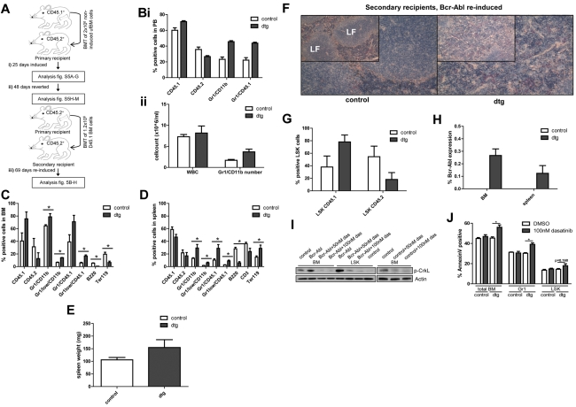
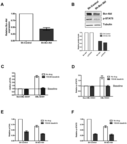
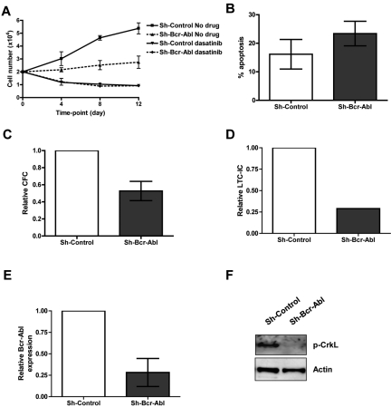
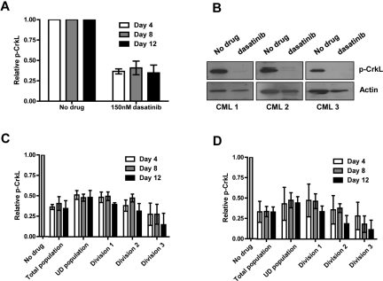
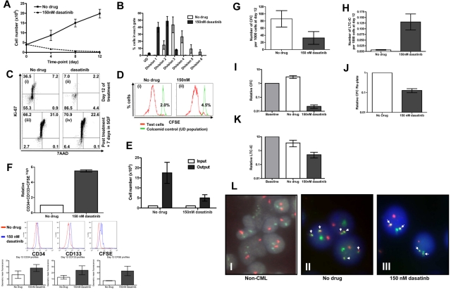
Recent evidence suggests chronic myeloid leukemia (CML) stem cells are insensitive to kinase inhibitors and responsible for minimal residual disease in treated patients. We investigated whether CML stem cells, in a transgenic mouse model of CML-like disease or derived from patients, are dependent on Bcr-Abl. In the transgenic model, after retransplantation, donor-derived CML stem cells in which Bcr-Abl expression had been induced and subsequently shut off were able to persist in vivo and reinitiate leukemia in secondary recipients on Bcr-Abl reexpression. Bcr-Abl knockdown in human CD34(+) CML cells cultured for 12 days in physiologic growth factors achieved partial inhibition of Bcr-Abl and downstream targets p-CrkL and p-STAT5, inhibition of proliferation and colony forming cells, but no reduction of input cells. The addition of dasatinib further inhibited p-CrkL and p-STAT5, yet only reduced input cells by 50%. Complete growth factor withdrawal plus dasatinib further reduced input cells to 10%; however, the surviving fraction was enriched for primitive leukemic cells capable of growth in a long-term culture-initiating cell assay and expansion on removal of dasatinib and addition of growth factors. Together, these data suggest that CML stem cell survival is Bcr-Abl kinase independent and suggest curative approaches in CML must focus on kinase-independent mechanisms of resistance.


Figures

Figure 1
Figure 1
CML stem cells retain transplantation and oncogenic capacity following Bcr-Abl reversion. (A) Irradiated CD45.2+ mice (8 Gy) transplanted using unfractionated (uf) CD45.1+ BM cells isolated from SCLtTA/Bcr-Abl dtg or wt controls (n = 12/12). (i) Bcr-Abl expression in recipient mice was controlled by absence (expression) or presence (no expression) of tetracycline in drinking water. Bcr-Abl expression was induced after transplantation for 25 days and the phenotype analyzed in dtg and controls (n = 3/3) by FACS of BM and spleen cells, spleen weight and histology, ratio of donor:host LSK cells, and Bcr-Abl expression. (ii) Bcr-Abl expression was reverted for 48 days and a cohort of remaining mice (n = 4/4) analyzed. (iii) BM cells from reverted dtg and controls were retransplanted using CD45.1+ FACS-sorted cells (n = 5/5). Bcr-Abl expression was reinduced and the phenotype was assessed 69 days after retransplantation. (Bi-ii) PB was analyzed in CD45.1+ retransplanted and reinduced mice 34 days after secondary transplantation. FACS of (C) BM and (D) spleen. Mean percentages of donor cells (CD45.1+), recipient cells (CD45.2+), granulocytes (Gr1+/CD11b+), immature granulocytes (Gr1low/CD11b+), donor granulocytes (Gr1+/CD45.1+), immature donor granulocytes (Gr1low/CD45.1+), B cells (B220+), T cells (CD3+), or erythroid cells (Ter119+). Spleen weights on (E) autopsy, (F) spleen histology, (G) donor:host ratio within BM LSK, and (H) Bcr-Abl expression in secondary recipients. (I) BM and LSK cells from 4-week induced dtg and control mice were cultured with either 50nM dasatinib, 100nM dasatinib, or DMSO for 48 hours. The level of p-CrkL was measured by Western blotting. (J) Apoptosis in total BM, Gr1, and LSK cells was assessed by annexin V staining in cells from 4-week induced dtg (n = 7) and control (n = 5) mice that were treated for 48 hours using 100nM dasatnib or DMSO. Histologic analyses of spleen were performed using NACE stain and are shown at magnifications of (insets) 10× and 20×. Shown is 1 representative spleen histology from each group. LF indicates lymphoid follicle. Data represented as mean ± SEM; *P < .05.
Figure 2
Figure 2
Bcr-Abl knockdown and dasatinib treatment of CML cells in physiologic growth factors. CD34+ CML cells were infected with sh-Bcr-Abl or sh-Control. (A) Forty-eight hours following second infection, cells were sorted for GFP and RNA, protein extracted, and relative expression of Bcr-Abl measured by Q-PCR (n = 5, P = .0004) and (B) Bcr-Abl and p-STAT5 by Western (n = 1). CD34+ non-CML (n = 3) and CML cells (n = 3) were cultured in SFM ± growth factors ± 150nM dasatinib. Twenty-four hours after treatment, (C) p-CrkL and (D) p-STAT5 were measured by flow cytometry. Lentivirally infected CD34+ cells (n = 3) were cultured in physiologic growth factors and treated ± 150nM dasatinib. Twenty-four hours after treatment, (E) p-CrkL and (F) p-STAT5 were measured in infected cells (gated on GFP) cells by flow cytometry.
Figure 3
Figure 3
Functional analysis of CML CD34+ cells following Bcr-Abl knockdown ± dasatinib. (A) Proliferation of GFP+ sorted cells treated ± 150nM dasatinib in physiologic growth factors over 12 days (n = 3). (B) Apoptosis measured in infected cells (gated on GFP) by annexin V and viaprobe (n = 2). (C) CFC (n = 4, P = .025) and (D) LTC-IC assays (n = 1). (E) Bcr-Abl levels were measured by Q-PCR on colonies from CFC assay (n = 7 colonies for sh-scrambled and 9 colonies for sh-Bcr-Abl). (F) The level of total p-CrkL was measured by Western blotting on pooled colonies from CFC assay (n = 1).
Figure 4
Figure 4
Complete Bcr-Abl inhibition is achieved by dasatinib within primitive subpopulations of CML cells. CFSE-stained CD34+ CML cells (n = 3) were cultured in SFM ± 150 or 1000nM dasatinib for 12 days. (A) The levels of total p-CrkL were measured by flow cytometry and (B) by Western blotting at day 12. p-CrkL was also measured within each cell division after treatment with either (C) 150 or (D) 1000nM dasatinib.
Figure 5
Figure 5
A primitive subset of CD34+ CML cells survives complete Bcr-Abl inhibition in growth factor–free medium for 12 days. (A) CD34+ CML cells (n = 3) were cultured in SFM ± 150nM and viable cell counts performed on days 4, 8, and 12, and (B) percentages of cells residing within each cell division determined by CFSE staining. After 12 days, cells were washed with PBS, added to CFC and LTC-IC assays, cultured in SFM plus high growth factor cocktail for 7 days. At day 7, (C) Ki-67/7AAD and (D) CFSE were measured by flow cytometry and (E) viable cell counts performed. (Ci) Cells at day 12 without drug treatment; (ii) cells at day 12 following dasatinb treatment; (iii) cells from (i) cultured for additional 7 days in SFM plus high growth cocktail; and (iv) cells from (ii) cultured for additional 7 days in SFM plus high growth factor cocktail. (Di) Cells from panel Ci were cultured for additional 7 days in SFM plus high growth factor cocktail, and (ii) cells from panel Cii were cultured for additional 7 days in SFM plus high growth factor cocktail. (F) The relative increase in CD34+CD133+CFSEhigh was calculated (n = 3, P = .001) and the numbers of (G) CFC (n = 7, P = .048) and (H) LTC-IC (n = 3, P = .025) per 1000 cells at day 12 were calculated. The changes in (I) CFC, (J) CFC replated (n = 2), and (K) LTC-IC numbers compared with baseline cells (1.0) were also calculated. (L) The presence of Bcr-Abl (denoted by the white arrows) was measured by D-FISH in the remaining CML cells following the 12 day time course. A representative profile from (i) non-CML CD34+ cells at baseline, (ii) untreated CD34+ CML cells and (iii) 150nM dasatinib-treated CD34+ cells is demonstrated.

Similar articles

Cited by

References

    1. Daley GQ, Van Etten RA, Baltimore D. Induction of chronic myelogenous leukemia in mice by the P210bcr/abl gene of the Philadelphia chromosome. Science. 1990;247(4944):824–830. - PubMed
    1. Koschmieder S, Gottgens B, Zhang P, et al. Inducible chronic phase of myeloid leukemia with expansion of hematopoietic stem cells in a transgenic model of BCR-ABL leukemogenesis. Blood. 2005;105(1):324–334. - PubMed
    1. Anderson K, Lutz C, van Delft FW, et al. Genetic variegation of clonal architecture and propagating cells in leukaemia. Nature. 2011;469(7330):356–361. - PubMed
    1. Notta F, Mullighan CG, Wang JC, et al. Evolution of human BCR-ABL1 lymphoblastic leukaemia-initiating cells. Nature. 2011;469(7330):362–367. - PubMed
    1. Druker BJ, Tamura S, Buchdunger E, et al. Effects of a selective inhibitor of the Abl tyrosine kinase on the growth of Bcr-Abl positive cells. Nat Med. 1996;2(5):561–566. - PubMed

Publication types

MeSH terms