. 2012 Mar;80(3):982-95.
doi: 10.1128/IAI.05918-11. Epub 2011 Dec 19.

Use of the chinchilla model for nasopharyngeal colonization to study gene expression by Moraxella catarrhalis

Affiliations

Use of the chinchilla model for nasopharyngeal colonization to study gene expression by Moraxella catarrhalis

Todd C Hoopman et al. Infect Immun. 2012 Mar.

Abstract

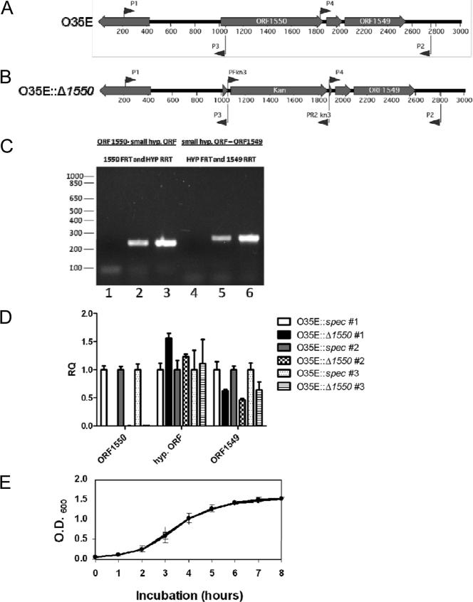
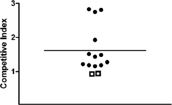
Young adult chinchillas were atraumatically inoculated with Moraxella catarrhalis via the nasal route. Detailed histopathologic examination of nasopharyngeal tissues isolated from these M. catarrhalis-infected animals revealed the presence of significant inflammation within the epithelium. Absence of similar histopathologic findings in sham-inoculated animals confirmed that M. catarrhalis was exposed to significant host-derived factors in this environment. Twenty-four hours after inoculation, viable M. catarrhalis organisms were recovered from the nasal cavity and nasopharynx of the animals in numbers sufficient for DNA microarray analysis. More than 100 M. catarrhalis genes were upregulated in vivo, including open reading frames (ORFs) encoding proteins that are involved in a truncated denitrification pathway or in the oxidative stress response, as well as several putative transcriptional regulators. Additionally, 200 M. catarrhalis genes were found to be downregulated when this bacterium was introduced into the nasopharynx. These downregulated genes included ORFs encoding several well-characterized M. catarrhalis surface proteins including Hag, McaP, and MchA1. Real-time reverse transcriptase PCR (RT-PCR) was utilized as a stringent control to validate the results of in vivo gene expression patterns as measured by DNA microarray analysis. Inactivation of one of the genes (MC ORF 1550) that was upregulated in vivo resulted in a decrease in the ability of M. catarrhalis to survive in the chinchilla nasopharynx over a 3-day period. This is the first evaluation of global transcriptome expression by M. catarrhalis cells in vivo.


Figures

Fig 1
Fig 1
Phenotypic comparison of strains O35E.118 and O35E.118.rpsL. (A) Growth of O35E.118 (closed circles) and O35E.118.rpsL (open circles) in BHI broth. (B) Outer membrane protein profiles of O35E.118 (lane 1) and O35E.118.rpsL (lane 2) as determined by SDS-PAGE and Coomassie blue staining.
Fig 2
Fig 2
Histopathologic analysis of the chinchilla response to intranasal inoculation with M. catarrhalis O35E.118.rpsL. (A and B) Histologic sections of the chinchilla nasal cavity in the sham-inoculated animal (A) and the M. catarrhalis strain O35E.118.rpsL-inoculated animal (B) 24 h following inoculation. These images depict the dorsal meatus from the region just caudal to the previously described level II in the chinchilla skull (32). Purulent exudate is evident within the nasal cavity overlying the intact epithelium (indicated by black arrows in B). (C and D) Higher magnification of the same mucosal regions in the sham-inoculated animal (C) and the M. catarrhalis O35E.118.rpsL-inoculated animal (D). Neutrophils are evident in migration between the epithelial cells in D (white arrow). Bars, 60 μm.
Fig 3
Fig 3
Relative expression levels of selected M. catarrhalis genes in the chinchilla nasopharynx as measured by real-time RT-PCR and DNA microarray methods. Expression of 26 selected genes in M. catarrhalis O35E.118.rpsL cells grown in broth or harvested from the chinchilla nasopharynx was measured by real-time RT-PCR analysis as described in Materials and Methods. The log2 of the fold difference in gene expression between in vivo and in vitro expression as determined by real-time RT-PCR (white columns) is plotted adjacent to the results obtained in DNA microarray analysis (black columns). MC ORF 1045 was chosen as an internal control for real-time RT-PCR analysis because this gene apparently did not alter its level of expression between in vitro and in vivo conditions as measured by DNA microarray analysis.
Fig 4
Fig 4
Characterization of M. catarrhalis O35EΔ1550. (A) Wild-type O35E locus containing MC ORF 1550. (B) Insertion of the Kan cartridge into a deletion within MC ORF 1550 in O35EΔ1550. (C) Reverse transcriptase PCR (RT-PCR)-based analysis of transcriptional linkage between selected ORFs downstream from MC ORF 1550 in the O35E::spec strain. The primer pair (Table 2) used for each experiment is specified at the top of the figure, as is the relevant intergenic region. Lanes: 1 and 4, samples from RT-PCR with RNA as the template but no added RT; 2 and 5, samples from RT-PCR with RNA as the template and RT added; 3 and 6, samples from RT-PCR with chromosomal DNA. Size position markers (in base pairs) are present on the left. A representative experiment is shown. (D) Real-time RT-PCR analysis of relative transcript levels for MC ORF 1550, the small hypothetical ORF (hyp. ORF), and MC ORF 1549 in O35EΔ1550 relative to the surrogate wild-type O35E::spec strain. Three independently isolated sets of RNA samples (#1, #2, and #3) were used for these experiments. Statistical analysis was performed using a one-sample t test with Bonferroni's correction. (E) Growth of the O35E::spec (closed circles) and O35EΔ1550 (open circles) strains in BHI broth. These data represent the mean from two independent experiments.
Fig 5
Fig 5
Competitive index values from mixed infections of the chinchilla nasopharynx. Mixtures of the O35E::spec surrogate wild-type strain and the O35EΔ1550 mutant were inoculated into the chinchilla nasopharynx. The input ratios varied from 0.70 to 0.90 in five independent experiments involving a total of 14 animals. After 72 h, the animals were euthanized and the relevant tissues were homogenized and plated on selective media to allow enumeration of each strain. The output ratios varied from 0.93 to 2.83. Competitive indices greater than 1.0 are indicated by black circles, and those less than 1.0 are indicated by open squares. The horizontal bar represents the mean of the competitive indices. A competitive index greater than 1.0 indicated that the wild-type surrogate strain outcompeted the mutant in that animal.

Similar articles

Cited by

References

    1. Aebi C, et al. 1998. Phenotypic effect of isogenic uspA1 and uspA2 mutations on Moraxella catarrhalis O35E. Infect. Immun. 66:3113–3119 - PMC - PubMed
    1. Agnew C, et al. 2011. Correlation of in situ mechanosensitive responses of the Moraxella catarrhalis adhesin UspA1 with fibronectin and receptor CEACAM1 binding. Proc. Natl. Acad. Sci. U. S. A. 108:15174–15178 - PMC - PubMed
    1. Armbruster CE, et al. 2010. Indirect pathogenicity of Haemophilus influenzae and Moraxella catarrhalis in polymicrobial otitis media occurs via interspecies quorum signaling. mBio 1:e00102–e00110 - PMC - PubMed
    1. Attia AS, Ram S, Rice PA, Hansen EJ. 2006. Binding of vitronectin by the Moraxella catarrhalis UspA2 protein interferes with late stages of the complement cascade. Infect. Immun. 74:1597–1611 - PMC - PubMed
    1. Bakaletz LO, Murwin DM, Billy JM. 1995. Adenovirus serotype 1 does not act synergistically with Moraxella (Branhamella) catarrhalis to induce otitis media in the chinchilla. Infect. Immun. 63:4188–4190 - PMC - PubMed

Publication types

Associated data