

Alveolar bone loss around incisors in Class I bidentoalveolar protrusion patients: a retrospective three-dimensional cone beam CT study
- PMID: 22184474
- PMCID: PMC3520391
- DOI: 10.1259/dmfr/30845402
Alveolar bone loss around incisors in Class I bidentoalveolar protrusion patients: a retrospective three-dimensional cone beam CT study
Abstract
Objectives: The aim of this study was to test the null hypothesis that there is no difference in the alveolar bone thickness, bone loss or incidence of fenestrations between upper and lower incisors in skeletal Class I bidentoalveolar protrusive patients before orthodontic treatment.
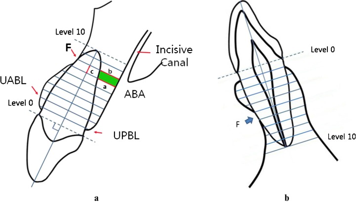
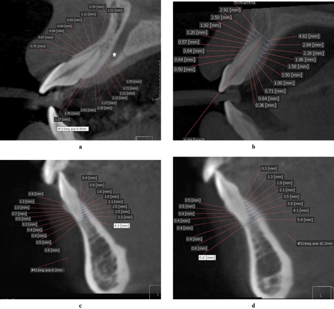
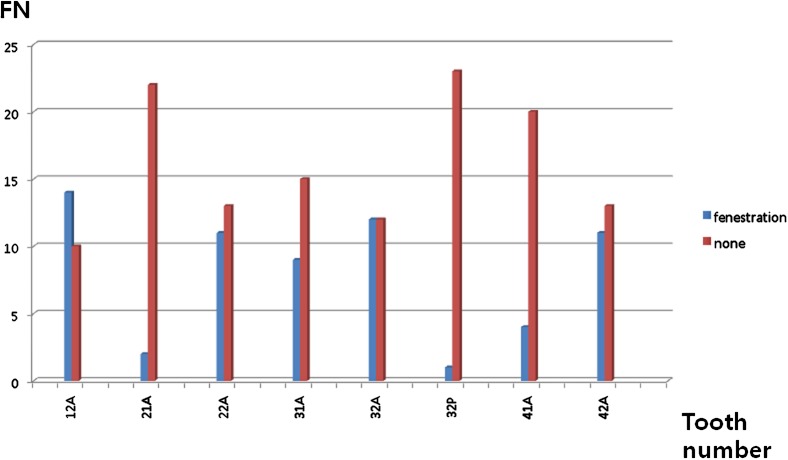
Methods: Three-dimensional (3D) cone beam CT (CBCT) images were taken of 24 patients from the Republic of Korea (17 females and 7 males). Reformatted CBCT images were used to measure labial and lingual alveolar bone thickness (ABT) of the 4 upper incisors and 4 lower incisors of the 24 patients (total n = 192 incisors) at every 1/10 of root length (Level 0, cementoenamel junction (CEJ) area; Level 10, root apex area) as well as alveolar bone area (ABA) and alveolar bone loss (%BL) rate to dental root length. The numbers of fenestration teeth were also tallied.
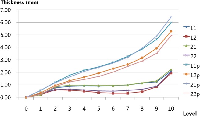
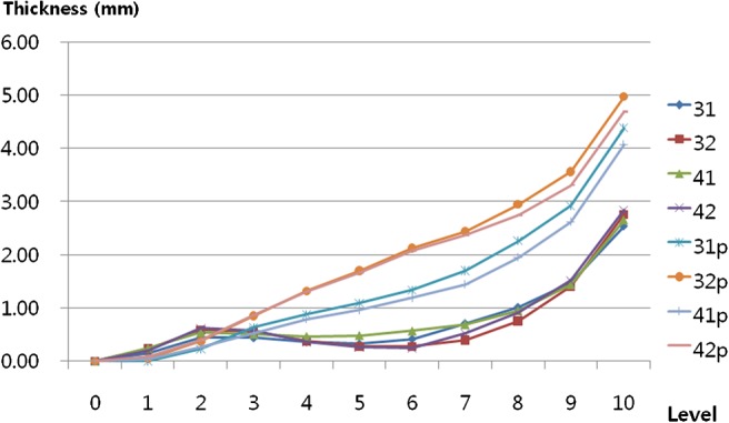
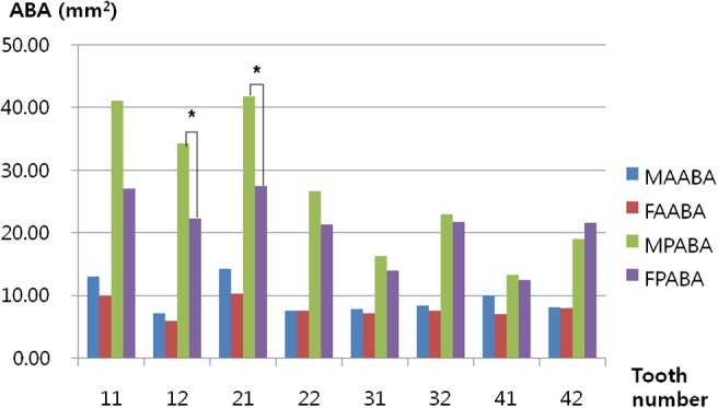
Results: All anterior teeth were supported by <1 mm of ABT on the labial surfaces up to root length Level 8. ABA was statistically greater on the lingual aspect than the labial aspect in lower incisors. The %BL was 26.98% in the lower labial region, 19.27% in upper labial aspect and most severe on the lower lingual plate 31.25% compared with the labial plate. There were no significant differences in %BL between subgroups when categorized by sex or age. Fenestrations were 1.37 times more frequent on lower incisors (37) than upper incisors (27).
Conclusion: The null hypothesis was rejected, confirming that incisor periodontal support is poor and alveolar bone loss is severe even prior to the start of orthodontic treatment. Careful diagnosis using 3D CBCT images is needed to avoid iatrogenic degeneration of periodontal support around anterior teeth, particularly in the lower lingual bone plate region.
Figures

Similar articles
-
CBCT-based assessment of apical root resorption and alveolar bone height following orthodontic treatment of Class I moderate crowding with labial vs. lingual fixed appliances in young adults: A randomized controlled trial.Int Orthod. 2025 Jun;23(2):100968. doi: 10.1016/j.ortho.2025.100968. Epub 2025 Jan 20. Int Orthod. 2025. PMID: 39837069 Clinical Trial.
-
Comparison of alveolar bone loss around incisors in normal occlusion samples and surgical skeletal class III patients.Angle Orthod. 2012 Jul;82(4):645-52. doi: 10.2319/070111-424.1. Epub 2011 Nov 30. Angle Orthod. 2012. PMID: 22129151 Free PMC article.
-
Three-dimensional assessment of periodontal support of lower incisors for skeletal Class II malocclusion undergoing presurgical orthodontic treatment with different vertical skeletal patterns.Prog Orthod. 2023 Dec 18;24(1):45. doi: 10.1186/s40510-023-00495-y. Prog Orthod. 2023. PMID: 38105288 Free PMC article.
-
Bone Remodeling during Orthodontic Movement of Lower Incisors-Narrative Review.Int J Environ Res Public Health. 2022 Nov 15;19(22):15002. doi: 10.3390/ijerph192215002. Int J Environ Res Public Health. 2022. PMID: 36429721 Free PMC article. Review.
-
Alveolar bone changes in maxillary and mandibular anterior teeth during orthodontic treatment: A systematic review and meta-analysis.Orthod Craniofac Res. 2021 May;24(2):165-179. doi: 10.1111/ocr.12421. Epub 2020 Sep 16. Orthod Craniofac Res. 2021. PMID: 32779352
Cited by
-
Alveolar bone thickness and fenestration of incisors in untreated Korean patients with skeletal class III malocclusion: A retrospective 3-dimensional cone-beam computed tomography study.Imaging Sci Dent. 2020 Mar;50(1):9-14. doi: 10.5624/isd.2020.50.1.9. Epub 2020 Mar 17. Imaging Sci Dent. 2020. PMID: 32206615 Free PMC article.
-
Performance of Cone Beam Computed Tomography Systems in Visualizing the Cortical Plate in 3D Image Reconstruction: An In Vitro Study.Open Dent J. 2018 Aug 31;12:586-595. doi: 10.2174/1874210601812010586. eCollection 2018. Open Dent J. 2018. PMID: 30288182 Free PMC article.
-
Bone Remodeling of Maxilla after Retraction of Incisors during Orthodontic Treatment with Extraction of Premolars Based on CBCT Study: A Systematic Review.J Clin Med. 2024 Mar 5;13(5):1503. doi: 10.3390/jcm13051503. J Clin Med. 2024. PMID: 38592367 Free PMC article. Review.
-
Changes of alveolar bone dehiscence and fenestration after augmented corticotomy-assisted orthodontic treatment: a CBCT evaluation.Prog Orthod. 2019 Feb 18;20(1):7. doi: 10.1186/s40510-019-0259-z. Prog Orthod. 2019. PMID: 30773604 Free PMC article.
-
Alveolar bone response to maxillary incisor retraction using stable skeletal structures as a reference.Angle Orthod. 2021 Jan 1;91(1):30-35. doi: 10.2319/022920-146.1. Angle Orthod. 2021. PMID: 33289780 Free PMC article.
References
-
- Handelman CS. The anterior alveolus: its importance in limiting orthodontic treatment and its influence on the occurrence or iatrogenic sequelae. Angle Orthod 1996;66:95–110 - PubMed
-
- Nelson PA, Årtun J. Alveolar bone loss of maxillary anterior teeth in adult orthodontic patients. Am J Orthod Dentofacial Orthop 1997;111:328–334 - PubMed
-
- Kim Y, Park JU, Kook YA. Alveolar bone loss around incisors in surgical skeletal Class III patients. Angle Orthod 2009;79:676–682 - PubMed
-
- Fuhrmann R. Three-dimensional interpretation of labiolingual bone width of the lower incisors. J Orofac Orthop 1996;57:168–185 - PubMed
-
- Nakajima K, Yamaguchi T, Maki K. Surgical orthodontic treatment for a patient with advanced periodontal disease: evaluation with electromyography and 3-dimensional cone-beam computed tomography. Am J Orthod Dentofacial Orthop 2009;136:450–459 - PubMed
MeSH terms
LinkOut - more resources
Full Text Sources
Other Literature Sources
Miscellaneous
