

Dural MALT lymphoma with disseminated disease
- PMID: 22184513
- PMCID: PMC3222263
- DOI: 10.4081/hr.2010.e10
Dural MALT lymphoma with disseminated disease
Abstract
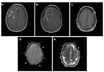
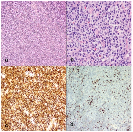
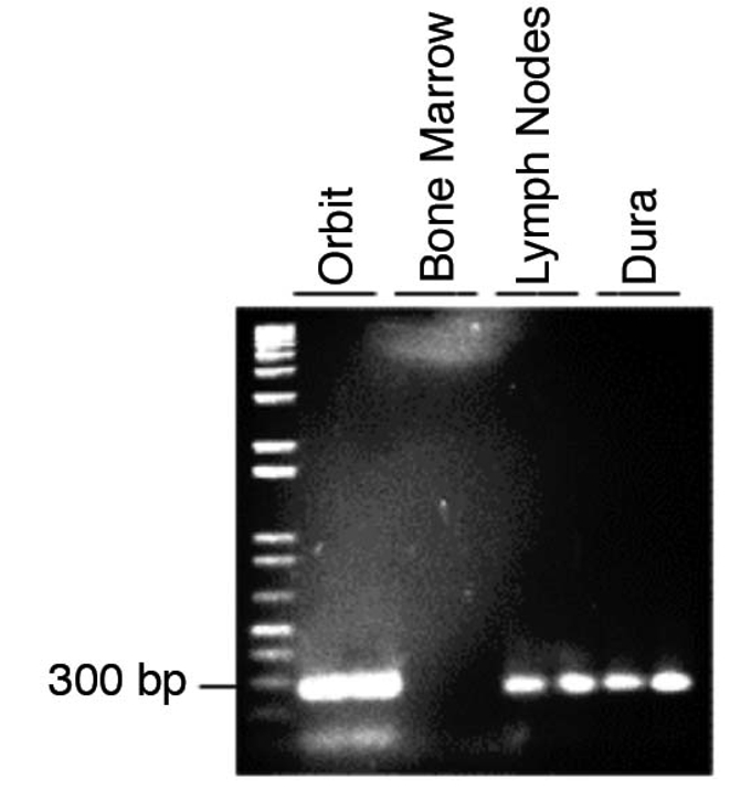
Central nervous system (CNS) lymphoma involving the dura mater is very rare and histologically is usually a subtype of non-Hodgkin's lymphoma (NHL) termed mucosa-associated lymphoid tissue (MALT) lymphoma. We present a case of a 46-year old woman with dural MALT lymphoma that was found to also involve a lacrimal gland, inguinal lymph nodes, and bone marrow. Magnetic resonance imaging of the brain showed an extra-axial enhancing mass approximately 6 cm in maximum diameter along the right frontotemporal convexity. Histopathology of the resected dural mass showed MALT lymphoma expressing CD20, CD52, CD19, and CD38. Molecular studies of the B-cell receptor heavy chain demonstrated monoclonality at the involved sites. The patient was treated with four cycles of fludarabine, mitoxantrone, and rituximab with complete remission. She had recurrence in the subcutaneous tissue of the back at 12 months but has remained free of intracranial disease for 31 months. A review of the literature reveals 57 cases of dural MALT lymphoma. Only 4 had extra-CNS involvement at presentation, and only 3 had local recurrence of the dural tumor. Because of the indolent behavior of this tumor, the intracranial portion can be treated conservatively after resection with or without chemotherapy. Deferral of brain radiation can be considered with close clinical and neuroimaging follow up.
Keywords: Chemotherapy; Dural lymphoma; MALT lymphoma; Meningioma.; Radiation.
Conflict of interest statement
Conflict of interest: the authors report no conflicts of interest.
Figures
Similar articles
-
Dural Follicular Lymphoma: Case Report and Literature Review.J Investig Med High Impact Case Rep. 2021 Jan-Dec;9:23247096211056768. doi: 10.1177/23247096211056768. J Investig Med High Impact Case Rep. 2021. PMID: 34844481 Free PMC article. Review.
-
Primary lymphoblastic B-cell lymphoma of the cranial dura mater: a case report and review of the literature.Leuk Lymphoma. 2005 Nov;46(11):1651-7. doi: 10.1080/10428190500215126. Leuk Lymphoma. 2005. PMID: 16334908
-
Extranodal Marginal Zone B-Cell Lymphoma of Mucosa-Associated Tissue Type Involving the Dura.Cancer Res Treat. 2016 Apr;48(2):859-63. doi: 10.4143/crt.2014.334. Epub 2015 Jul 17. Cancer Res Treat. 2016. PMID: 26194368 Free PMC article.
-
Mucosa-associated lymphoid tissue lymphoma of the dura mimicking meningioma: A case report.Surg Neurol Int. 2025 Feb 28;16:63. doi: 10.25259/SNI_902_2023. eCollection 2025. Surg Neurol Int. 2025. PMID: 40041086 Free PMC article.
-
Extranodal marginal zone lymphoma of mucosa-associated lymphoid tissue of the dura mimicking meningioma: a case report and literature review.Folia Neuropathol. 2024;62(1):102-107. doi: 10.5114/fn.2024.135291. Folia Neuropathol. 2024. PMID: 38741437 Review.
Cited by
-
Rare case of cerebral MALToma presenting with stroke-like symptoms and seizures.BMJ Case Rep. 2013 Apr 22;2013:bcr2012008494. doi: 10.1136/bcr-2012-008494. BMJ Case Rep. 2013. PMID: 23608841 Free PMC article.
-
Primary anaplastic large cell lymphoma in the dura of the brain: case report and prediction of a favorable prognosis.Int J Clin Exp Pathol. 2013 Jul 15;6(8):1643-51. Print 2013. Int J Clin Exp Pathol. 2013. PMID: 23923083 Free PMC article.
-
An international multicenter retrospective analysis of patients with extranodal marginal zone lymphoma and histologically confirmed central nervous system and dural involvement.Cancer Med. 2020 Jan;9(2):663-670. doi: 10.1002/cam4.2732. Epub 2019 Dec 5. Cancer Med. 2020. PMID: 31808316 Free PMC article.
-
Dural Follicular Lymphoma: Case Report and Literature Review.J Investig Med High Impact Case Rep. 2021 Jan-Dec;9:23247096211056768. doi: 10.1177/23247096211056768. J Investig Med High Impact Case Rep. 2021. PMID: 34844481 Free PMC article. Review.
-
A Rare Case of Composite Dural Extranodal Marginal Zone Lymphoma and Chronic Lymphocytic Leukemia/Small Lymphocytic Lymphoma.Front Neurol. 2018 Apr 24;9:267. doi: 10.3389/fneur.2018.00267. eCollection 2018. Front Neurol. 2018. PMID: 29740389 Free PMC article. Review.
References
-
- Commins DL. Pathology of primary central nervous system lymphoma. Neurosurg Focus. 2006;21:E2. - PubMed
-
- Gleissner B, Chamberlain M. Treatment of CNS dissemination in systemic lymphoma. J Neurooncol. 2007;84:107–17. - PubMed
-
- Low I, Allen J. Low-grade follicular lymphoma in the dura: rare mimic of meningioma. Neuropathology. 2006;26:564–8. - PubMed
-
- Bierman P, Giglio P. Diagnosis and treatment of central nervous system involvement in non-Hodgkin's lymphoma. Hematol Oncol Clin North Am. 19:597–609. - PubMed
-
- Iwamoto FM, Abrey LE. Primary dural lymphomas: a review. Neurosurg Focus. 2006;21:E5. - PubMed
Publication types
LinkOut - more resources
Full Text Sources
Research Materials
