. 2010 Jan 26;2(1):e2.
doi: 10.4081/hr.2010.e2. Epub 2010 Apr 13.

Pathological basis of symptoms and crises in sickle cell disorder: implications for counseling and psychotherapy

Affiliations

Pathological basis of symptoms and crises in sickle cell disorder: implications for counseling and psychotherapy

Oluwatoyin Olatundun Ilesanmi. Hematol Rep. .

Abstract

Sickle Cell Disorder (SCD) is a congenital hemoglobinopathy. There is little in literature regarding the psychological variables affecting individuals living with SCD and all of the significant people around them. There are also limited numbers of trained clinical psychologists and genetic counselors to cater for the psychotherapeutic needs of individuals living with SCD. Even among those who have been trained, only a few might have fully grasped the complexities of the disease pathology.Early understanding of its pathological nature, sources, types, complications, pathophysiological basis, and clinical severity of symptoms among clinical psychologists, genetic counselors and psychotherapists, as well as general medical practitioners, could guide them in providing holistic care for dealing with and reducing pain among individuals living with SCD. It could allow risk-based counseling for families and individuals. It could also justify the early use of disease-modifying or curative interventions, such as hydroxyurea (HU), chronic transfusions (CTs), or stem-cell transplantation (SCT) by general medical practitioners. Hence, the need for this paper on the pathophysiology of SCD.

Keywords: counseling and psychotherapy.; implications; pathology; sickle cell disorder.


Figures
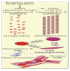
Figure 1
Figure 1
Molecular and cellular changes of hemoglobin S. Legend: β6Glu⇒Val. Deoxy Hb S polymer forms with low O2, Causes irreversibly sickled cells. Under a variety of circumstances, different organs are susceptible: esp. bone, spleen, and lung.
Figure 2
Figure 2
Oxy versus deoxyhemoglobin. Legend: α = Alpha globins; β= Beta globins.
Figure 3
Figure 3
Red cell morphology. Legend: RBC: red blood cells i) Endothelial activation; ii) WBC adhesion; iii) RBC-WBC interactions; iv) vasoocclusion. Lumen obstruction results from interaction b/w WBC, RBC, platelets, plasma proteins & vascular endothelium.
Figure 4
Figure 4
RBC membrane structure. Legend: I = Integral Proteins; P = Peripheral Proteins; hemoglobin occupies 33% of the RBC volume and 90–95% of the dry weight; 65% of the hemoglobin synthesis occurs in the nucleated stages of RBC maturation and 35% during the reticulocyte stage; normal hemoglobin consists of 4 heme groups, which contain a protoporphyrin ring plus iron, and globin, which is a tetramer of 2 pairs of polypeptide chains.
Figure 5
Figure 5
α= Alpha Chains. The α-globin genes are on chromosome 16. β=Beta Chains. The β-globin genes are on chromosome 11. Fe2+ = Ferrous indicates a bivalent iron compound (+2 oxidation state), as opposed to ferric, which indicates a trivalent iron compound (+3 oxidation state). Heme = is a prosthetic group that consists of an iron atom contained in the center of a large heterocyclic organic ring called a porphyrin. Not all porphyrins contain iron, but a substantial fraction of porphyrin-containing metalloproteins have heme as their prosthetic group; these are known as hemoproteins.
Figure 6
Figure 6
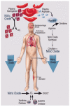
Depletion of nitric oxide in sickle cell anemia. Legend: Sickle cell disease Hemolysis Arginine ↓ NO ↓. During intravascular hemolysis, hemoglobin is released into the plasma where it is normally cleared by the hemoglobin scavengers haptoglobin, CD163, and hemopexin. Haptoglobin-hemoglobin complexes bind to CD163 on the surface of macrophages/ monocytes initiating endocytosis and degradation of the complex. Hemoglobin also releases ferric heme on oxidation, which is bound by hemopexin and degraded by hepatocytes in the liver. Excessive hemolysis saturates and depletes these hemoglobin removal systems and leads to a build-up of hemoglobin and heme in the plasma. Plasma hemoglobin and heme mediate direct proinflammatory, proliferative, and pro-oxidant effects on vessel endothelial cells. NO is normally generated from L-arginine in vessel endothelial cells by the enzyme nitric oxide synthase (NOS). NO maintains smooth muscle relaxation and inhibits platelet activation and aggregation, thereby regulating vessel tone and promoting organ system homeostasis. During intravascular hemolysis, NO availability can be severely limited by its reaction with oxyhemoglobin (NO scavenging) and by the breakdown of the substrate for NO synthesis, L-arginine, by the red cell enzyme arginase, despite elevated levels of NOS (decreased NO synthesis). NO depletion results in decreased activation of guanylate cyclase, an enzyme required for the generation of cyclic guanine mono-phosphate (cGMP). Decreased cGMP levels disrupt regulation of smooth muscle tone resulting in dystonias, including systemic and pulmonary hypertension, erectile dysfunction, dysphagia, and abdominal pain. Decreased cGMP levels through the depletion of NO can also lead to platelet activation and aggregation, promoting clot formation. GTP indicates guanosine 5′-triphosphate. (Modified from JAMA 2009).
Figure 7
Figure 7
Ballas figure on molecular mechanisms of pain.
Figure 8
Figure 8
The Coagulation Cascade. Legend: this is the physiological coagulation cascade used to describe a very complex step-by-step process that occurs in the body (in vivo) when a blood vessel is injured. In the cascade, solid arrows indicate activation of a coagulation factor by the factor above it in the cascade, dotted arrows indicate activation of factors V, VIII, XI and XIII by thrombin. The intrinsic cascade (which has less in vivo significance in normal physiological circumstances than the extrinsic cascade) is initiated when contact is made between blood and exposed negatively charged surfaces. The extrinsic pathway is initiated upon vascular injury which leads to exposure of tissue factor, TF (also identified as factor III), a subendothelial cell-surface glycoprotein that binds phospholipid. The green dotted arrow represents a point of cross-over between the extrinsic and intrinsic pathways. The two pathways converge at the activation of factor X to Xa. Factor Xa has a role in the further activation of factor VII to VIIa as depicted by the green arrow. Active factor Xa hydrolyzes and activates prothrombin to thrombin. Thrombin can then activate factors XI, VIII and V furthering the cascade. Ultimately the role of thrombin is to convert fribrinogen to fibrin and to activate factor XIII to XIIIa. Factor XIIIa (also termed transglutaminase) cross-links fibrin polymers solidifying the clot. HMWK = high molecular weight kininogen. PK = prekallikrein. PL = phospholipid.
Figure 9
Figure 9
Extravascular destruction of RBCs. Legend: Extravascular hemolysis occurs when damaged or abnormal RBCs are cleared from the circulation by cells of the spleen, liver, and bone marrow similar to the process by which senescent RBCs are removed. The spleen usually contributes to hemolysis by destroying mildly abnormal RBCs or cells coated with warm antibodies. An enlarged spleen may sequester even normal RBCs. Severely abnormal RBCs or RBCs coated with cold antibodies or complement (C3) are destroyed within the circulation and in the liver, which (because of its large blood flow) can remove damaged cells efficiently.
Figure 10
Figure 10
Intravascular destruction of RBCs. Intravascular hemolysis usually occurs when the cell membrane has been severely damaged by any of a number of different mechanisms, including autoimmune phenomena, direct trauma (e.g., march hemoglobinuria), shear stress (e.g., defective mechanical heart valves), and toxins (e.g., clostridial toxins, venomous snake bite). It results in hemoglobinemia when the amount of Hb released into plasma exceeds the Hb-binding capacity of the plasma-binding protein haptoglobin, a globulin normally present in concentrations of about 1.0 g/L in plasma. With hemoglobinemia, unbound Hb dimers are filtered into the urine and reabsorbed by renal tubular cells; hemoglobinuria results when reabsorptive capacity is exceeded. Iron is embedded in hemosiderin within the tubular cells; some of the iron is assimilated for reutilization and some reaches the urine when the tubular cells slough.
Figure 11
Figure 11
Avascular necrosis (osteonecrosis) of bone. Avascular necrosis of the hip occurs when blood flow to the top portion of the thighbone (femur) is interrupted. The affected portion of the bone consists of the head (the ball-shaped piece of bone that fits into the socket of the hip) and neck (the portion of the thighbone just below the head). When it's deprived of blood, this part of the bone begins to “die,” breaking down and causing the cartilage on top of it to collapse.

Similar articles

Cited by

References

    1. Embury SH, Hebbel RP, Mohandas N, Steinberg MH, editors. Basic Principles and Clinical Picture. New York: Raven Press; 1994. Sickle cell disease; pp. 311–311.
    1. Serjeant GR. 3rd ed. New York: Oxford University Press; 2001. Sickle cell disease.
    1. Ballas SK. Progress in Pain Research and Management. Vol. 11. Seattle, WA: IASP Press; 1998. Sickle Cell Pain.
    1. Benjamin LJ, Dampier CD, Jacox A, et al. American Pain Society Clinical Practice Guidelines Series No. 1. Glenview, IL: 1999. Guideline for the Management of Acute and Chronic Pain in Sickle cell disease.
    1. Benjamin LJ. Nature and treatment of the acute painful episode in sickle cell disease. In: Steinberg MH, et al., editors. Disorders of Hemoglobin: Genetics, Pathophysiology, and Clinical Management. Cambridge: 2001. pp. 671–710.