

Application of in vivo micro-computed tomography in the temporal characterisation of subchondral bone architecture in a rat model of low-dose monosodium iodoacetate-induced osteoarthritis
- PMID: 22185204
- PMCID: PMC3334663
- DOI: 10.1186/ar3543
Application of in vivo micro-computed tomography in the temporal characterisation of subchondral bone architecture in a rat model of low-dose monosodium iodoacetate-induced osteoarthritis
Abstract
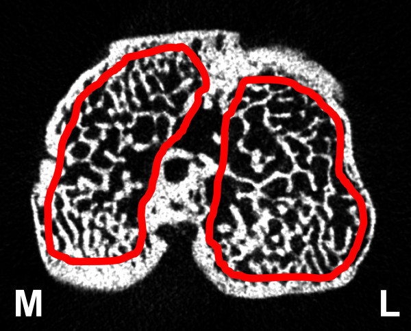
Introduction: Osteoarthritis (OA) is a complex, multifactorial joint disease affecting both the cartilage and the subchondral bone. Animal models of OA aid in the understanding of the pathogenesis of OA and testing suitable drugs for OA treatment. In this study we characterized the temporal changes in the tibial subchondral bone architecture in a rat model of low-dose monosodium iodoacetate (MIA)-induced OA using in vivo micro-computed tomography (CT).
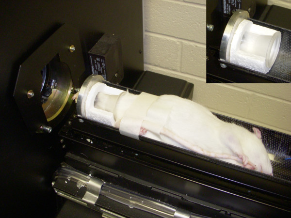
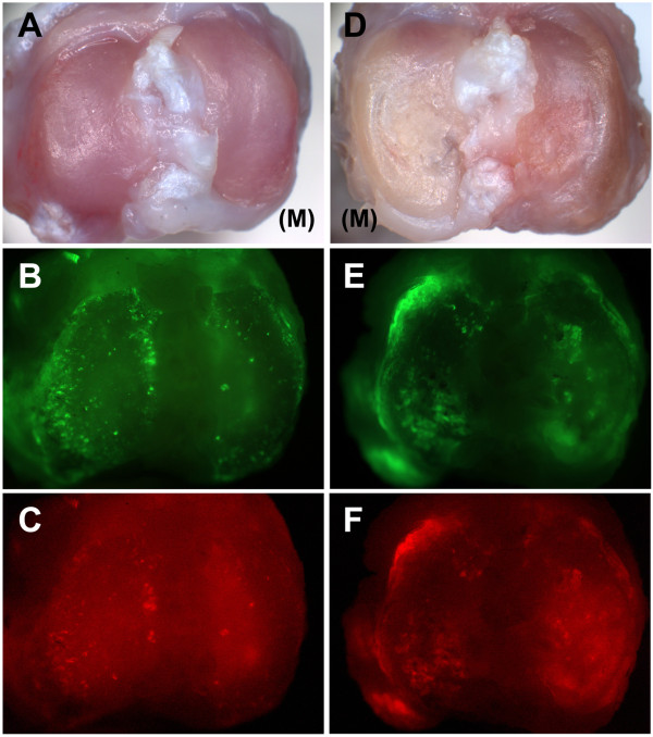
Methods: Male Wistar rats received a single intra-articular injection of low-dose MIA (0.2 mg) in the right knee joint and sterile saline in the left knee joint. The animals were scanned in vivo by micro-CT at two, six, and ten weeks post-injection, analogous to early, intermediate, and advanced stages of OA, to assess architectural changes in the tibial subchondral bone. The articular cartilage changes in the tibiae were assessed macroscopically and histologically at ten weeks post-injection.
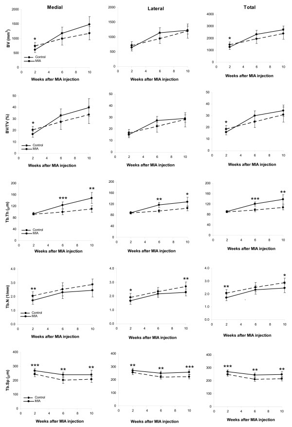
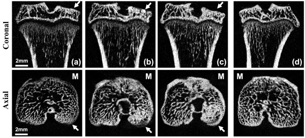
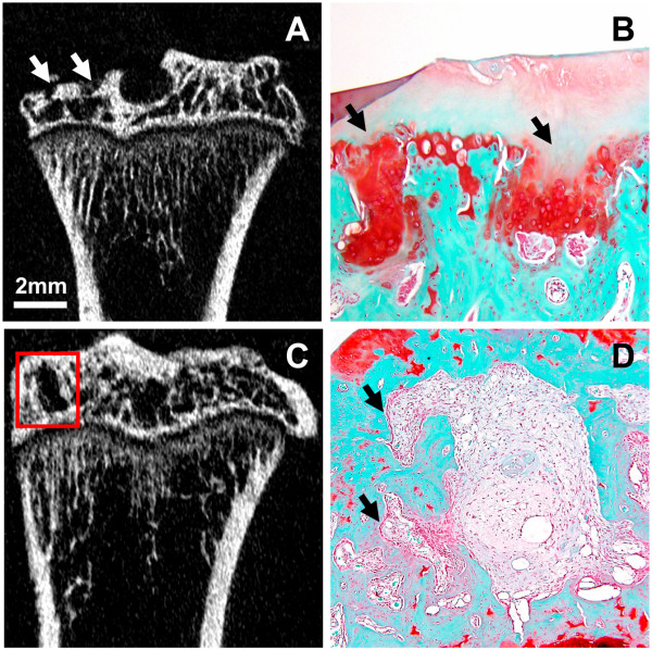
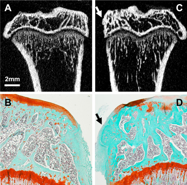
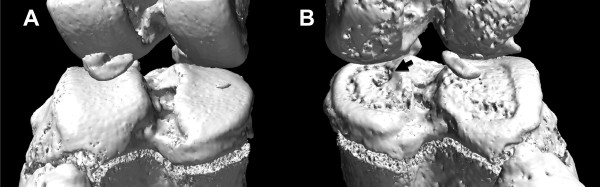
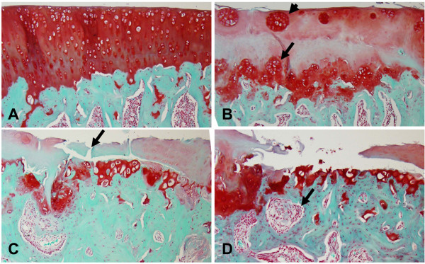
Results: Interestingly, tibiae of the MIA-injected knees showed significant bone loss at two weeks, followed by increased trabecular thickness and separation at six and ten weeks. The trabecular number was decreased at all time points compared to control tibiae. The tibial subchondral plate thickness of the MIA-injected knee was increased at two and six weeks and the plate porosity was increased at all time points compared to control. At ten weeks, histology revealed loss of proteoglycans, chondrocyte necrosis, chondrocyte clusters, cartilage fibrillation, and delamination in the MIA-injected tibiae, whereas the control tibiae showed no changes. Micro-CT images and histology showed the presence of subchondral bone sclerosis, cysts, and osteophytes.
Conclusions: These findings demonstrate that the low-dose MIA rat model closely mimics the pathological features of progressive human OA. The low-dose MIA rat model is therefore suitable to study the effect of therapeutic drugs on cartilage and bone in a non-trauma model of OA. In vivo micro-CT is a non-destructive imaging technique that can track structural changes in the tibial subchondral bone in this animal model, and could also be used to track changes in bone in preclinical drug intervention studies for OA treatments.
Figures
Similar articles
-
Pre-emptive, early, and delayed alendronate treatment in a rat model of knee osteoarthritis: effect on subchondral trabecular bone microarchitecture and cartilage degradation of the tibia, bone/cartilage turnover, and joint discomfort.Osteoarthritis Cartilage. 2013 Oct;21(10):1595-604. doi: 10.1016/j.joca.2013.06.020. Epub 2013 Jul 1. Osteoarthritis Cartilage. 2013. PMID: 23827368
-
In vivo micro computed tomography of subchondral bone in the rat after intra-articular administration of monosodium iodoacetate.Contemp Top Lab Anim Sci. 2004 Jan;43(1):39-43. Contemp Top Lab Anim Sci. 2004. PMID: 14984289
-
Mono-iodoacetate-induced histologic changes in subchondral bone and articular cartilage of rat femorotibial joints: an animal model of osteoarthritis.Toxicol Pathol. 2003 Nov-Dec;31(6):619-24. doi: 10.1080/01926230390241800. Toxicol Pathol. 2003. PMID: 14585729
-
Temporal progression of subchondral bone alterations in OA models involving induction of compromised meniscus integrity in mice and rats: A scoping review.Osteoarthritis Cartilage. 2024 Oct;32(10):1220-1234. doi: 10.1016/j.joca.2024.06.002. Epub 2024 Jun 12. Osteoarthritis Cartilage. 2024. PMID: 38876436
-
Advancements in Subchondral Bone Biomechanics: Insights from Computed Tomography and Micro-Computed Tomography Imaging in Equine Models.Curr Osteoporos Rep. 2024 Dec;22(6):544-552. doi: 10.1007/s11914-024-00886-y. Epub 2024 Sep 14. Curr Osteoporos Rep. 2024. PMID: 39276168 Free PMC article. Review.
Cited by
-
Chondroprotective effects of platelet lysate towards monoiodoacetate-induced arthritis by suppression of TNF-α-induced activation of NF-ĸB pathway in chondrocytes.Aging (Albany NY). 2019 May 14;11(9):2797-2811. doi: 10.18632/aging.101952. Aging (Albany NY). 2019. PMID: 31089001 Free PMC article.
-
Progression of cartilage degradation, bone resorption and pain in rat temporomandibular joint osteoarthritis induced by injection of iodoacetate.PLoS One. 2012;7(9):e45036. doi: 10.1371/journal.pone.0045036. Epub 2012 Sep 11. PLoS One. 2012. PMID: 22984604 Free PMC article.
-
Quantitative histological grading methods to assess subchondral bone and synovium changes subsequent to medial meniscus transection in the rat.Connect Tissue Res. 2017 May-Jul;58(3-4):373-385. doi: 10.1080/03008207.2016.1251425. Epub 2016 Oct 31. Connect Tissue Res. 2017. PMID: 27797605 Free PMC article.
-
SPHARM-PDM based image preprocessing pipeline for quantitative morphometric analysis (QMA) for in situ joint assessment in rabbit and rat models.Sci Rep. 2022 Jan 21;12(1):1113. doi: 10.1038/s41598-021-04542-8. Sci Rep. 2022. PMID: 35064147 Free PMC article.
-
Histology, glycosaminoglycan level and cartilage stiffness in monoiodoacetate-induced osteoarthritis: comparative analysis with anterior cruciate ligament transection in rat model and human osteoarthritis.Int J Med Sci. 2013 Dec 21;11(1):97-105. doi: 10.7150/ijms.6964. eCollection 2014. Int J Med Sci. 2013. PMID: 24396291 Free PMC article.
References
-
- Bailey AJ, Mansell JP, Sims TJ, Banse X. Biochemical and mechanical properties of subchondral bone in osteoarthritis. Biorheology. 2004;41:349–358. - PubMed
-
- Kalbhen DA. Chemical model of osteoarthritis--a pharmacological evaluation. J Rheumatol. 1987;14(Spec No):130–131. - PubMed
-
- Janusz MJ, Hookfin EB, Heitmeyer SA, Woessner JF, Freemont AJ, Hoyland JA, Brown KK, Hsieh LC, Almstead NG, De B, Natchus MG, Pikul S, Taiwo YO. Moderation of iodoacetate-induced experimental osteoarthritis in rats by matrix metalloproteinase inhibitors. Osteoarthritis Cartilage. 2001;9:751–760. doi: 10.1053/joca.2001.0472. - DOI - PubMed
Publication types
MeSH terms
Substances
LinkOut - more resources
Full Text Sources
Medical
