. 2011 Dec 20:12:129.
doi: 10.1186/1471-2202-12-129.

The nestin-expressing and non-expressing neurons in rat basal forebrain display different electrophysiological properties and project to hippocampus

Affiliations

The nestin-expressing and non-expressing neurons in rat basal forebrain display different electrophysiological properties and project to hippocampus

Jianhua Zhu et al. BMC Neurosci. .

Abstract

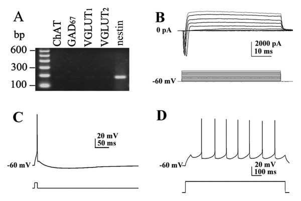
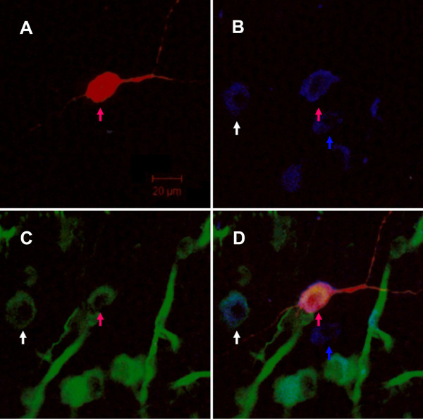
Background: Nestin-immunoreactive (nestin-ir) neurons have been identified in the medial septal/diagonal band complex (MS/DBB) of adult rat and human, but the significance of nestin expression in functional neurons is not clear. This study investigated electrophysiological properties and neurochemical phenotypes of nestin-expressing (nestin+) neurons using whole-cell recording combined with single-cell RT-PCR to explore the significance of nestin expression in functional MS/DBB neurons. The retrograde labelling and immunofluorescence were used to investigate the nestin+ neuron related circuit in the septo-hippocampal pathway.

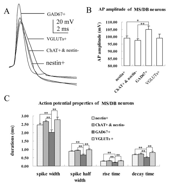
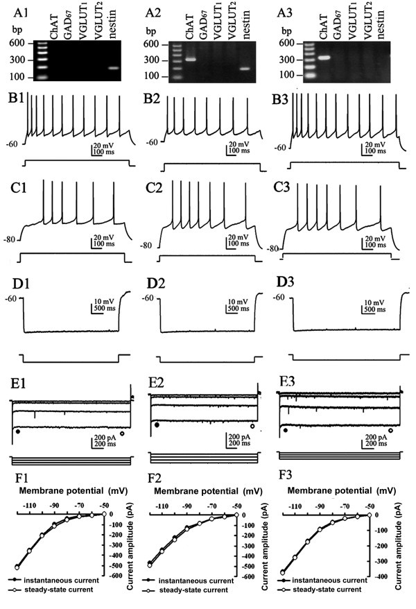
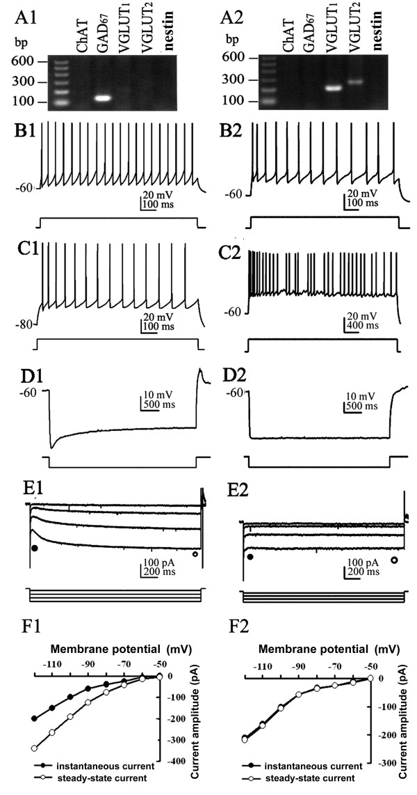
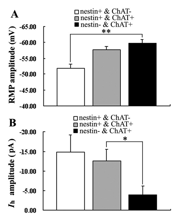
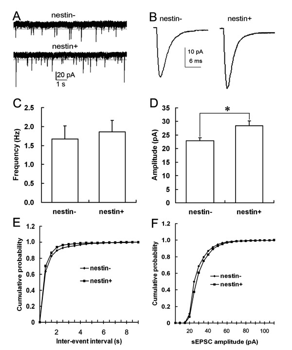
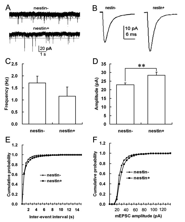
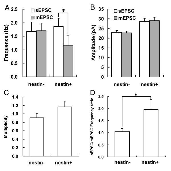
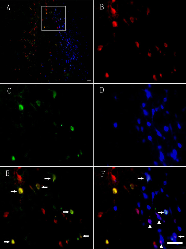
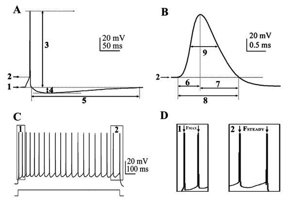
Results: The results of single-cell RT-PCR showed that 87.5% (35/40) of nestin+ cells expressed choline acetyltransferase mRNA (ChAT+), only 44.3% (35/79) of ChAT+ cells expressed nestin mRNA. Furthermore, none of the nestin+ cells expressed glutamic acid decarboxylases 67 (GAD(67)) or vesicular glutamate transporters (VGLUT) mRNA. All of the recorded nestin+ cells were excitable and demonstrated slow-firing properties, which were distinctive from those of GAD(67) or VGLUT mRNA-positive neurons. These results show that the MS/DBB cholinergic neurons could be divided into nestin-expressing cholinergic neurons (NEChs) and nestin non-expressing cholinergic neurons (NNChs). Interestingly, NEChs had higher excitability and received stronger spontaneous excitatory synaptic inputs than NNChs. Retrograde labelling combined with choline acetyltransferase and nestin immunofluorescence showed that both of the NEChs and NNChs projected to hippocampus.

Conclusions: These results suggest that there are two parallel cholinergic septo-hippocampal pathways that may have different functions. The significance of nestin expressing in functional neurons has been discussed.


Figures

Figure 1
Figure 1
Nestin mRNA-positive cells in MS/DBB are functional neurons. A. Agarose gel analysis of the sc-RT-PCR products obtained from a single MS/DBB cell. The only PCR-generated fragment was nestin. B. Whole cell current of the same cell depolarized from -60 mV to 10 mV in voltage-clamp mode showed characteristic of functional neurons. C. The cell shows typical neural action potential when depolarized to the threshold potential. D. A sustained depolarizing current (1000 ms) elicits a train of repetitive action potentials. The membrane responses were elicited using positive currents of 0.2 nA from -60 mV in panel C and D.
Figure 2
Figure 2
Comparison of action potential properties of MS/DBB neurons. A. Representative action potentials from all cell types (superimposed) show the differences in spike shape and width among the four cell types. GAD67+ neurons have the narrowest action potentials, where as VGLUT+ neurons have the broadest action potentials. B. Histogram of action potential amplitude of all cell types. C. Histogram of spike parameters of all cell types. * P < 0.05;** P < 0.01.
Figure 3
Figure 3
Electrophysiological properties of nestin+ and/or ChAT+ neurons. A1-F1: Nestin+ & ChAT- neurons, A2-F2: Nestin+ & ChAT+ neurons, A3-F3: Nestin- & ChAT+ neurons. A1-3: Agarose gel analysis of the sc-RT-PCR products to identify chemical phenotypes of recorded neurons. B1-3 and C1-3: In current-clamp mode, membrane responses of the same MS/DBB neuron to depolarizing current pulses (0.2 nA) applied from membrane potential of -60 mV (B1-3) or -80 mV (C1-3). All neurons display slow-firing activity. D1-3: Injection of a hyperpolarizing current pulse from -60 mV, no depolarizing sag and rebound firing were found in each kind of neuron. E1-3: Hyperpolarizing voltage steps applied from a holding potential of -50 mV showed absence of conspicuous Ih in all groups of neurons. F1-3: I-V plots of instantaneous (filled circle) and steady-state (open circle) current derived from the data in E1-3. The Ih of nestin+ & ChAT+ neuron was mildly larger than that of nestin- & ChAT+ neuron (E2, E3, F2, F3).
Figure 4
Figure 4
Electrophysiological properties of GAD67+ and VGLUT+ neurons. A1-F1: GAD67+ neuron presents electrophysiological properties of fast-firing neuron. A2-F2: VGLUT+ neuron presents electrophysiological properties of cluster-firing neuron. A1-2: Agarose gel analysis of the sc-RT-PCR products to identify chemical phenotypes of recorded neurons. B1 and C1: Membrane responses of the same MS/DBB neuron to injection of depolarizing current pulses applied from a membrane potential of -60 mV (B1) or -80 mV (C1). GAD67+ neuron displays fast-firing activity. B2 and C2: Membrane responses of the VGLUT+ neuron to injection of depolarizing current pulses (0.2 nA) applied from a membrane potential of -60 mV, prolonged depolarization current (4s in duration) elicits cluster-firing separated by subthreshold membrane oscillations (C2). D1-2: In current-clamp mode, the response to injections of hyperpolarizing current pulse from -60 mV, note profound depolarizing sag in GAD67+ neuron, but no rebound firing action potential (D1). E1-2: Currents recorded in voltage-clamp mode evoked by a series of hyperpolarizing voltage steps applied from a holding potential of -50 mV. Notice the profound depolarizing amplitude inward current in GAD67+ neuron, as shown by the differences between the amplitudes of the instantaneous current (filled circle) and steady-state current (open circle). F1-2: Instantaneous and steady-state I-V plots derived from the data in E1-2.
Figure 5
Figure 5
Comparison of RMP and Ih amplitude of nestin+ and/or ChAT+ neurons in MS/DBB. * P < 0.05; ** P < 0.01.
Figure 6
Figure 6
Comparison of sEPSCs of MS/DBB nestin- and nestin+ cholinergic neurons. A. Consecutive traces of sEPSCs recorded from MS/DBB nestin- and nestin+ cholinergic neurons. B. Average sEPSCs of nestin- and nestin+ cholinergic neurons. C. Comparison of the sEPSCs frequencies of MS/DBB nestin- and nestin+ cholinergic neurons. D. Comparison of the sEPSCs amplitudes of MS/DBB nestin- and nestin+ cholinergic neurons (*: P < 0.05). E. Cumulative probability distribution of sEPSCs inter-event intervals of nestin- and nestin+ cholinergic neurons in MS/DBB: the curve of nestin+ cholinergic neurons was on the left of the curve for nestin- cholinergic neurons (K-S Z = 2.644, P < 0.01); F. Cumulative probability distribution of sEPSCs amplitudes of nestin- and nestin+ cholinergic neurons in MS/DBB: the curve of nestin+ cholinergic neurons was on the right of the curve for nestin- cholinergic neurons (K-S Z = 4.549, P < 0.01).
Figure 7
Figure 7
Comparison of mEPSCs of MS/DBB nestin- and nestin+ cholinergic neurons. A. Consecutive traces recorded from MS/DBB nestin- and nestin+ cholinergic neurons. B. Average mEPSCs of nestin- and nestin+ cholinergic neurons. C. Comparison of the mEPSCs frequencies of MS/DBB nestin- and nestin+ cholinergic neurons. D. Comparison of the mEPSCs amplitudes of MS/DBB nestin- and nestin+ cholinergic neurons (**: P < 0.01). E. Cumulative probability distribution of mEPSCs inter-event intervals of nestin- and nestin+ cholinergic neurons in MS/DBB: the curve of nestin+ cholinergic neurons was on the right of that of nestin- cholinergic neurons (K-S Z = 1.717, P < 0.01). F. Cumulative probability distribution of mEPSCs amplitudes of nestin- and nestin+ cholinergic neurons in MS/DBB: the curve of nestin+ cholinergic neurons was on the right of that of the nestin- cholinergic neurons (K-S Z = 8.217, P < 0.01).
Figure 8
Figure 8
Comparison of sEPSCs and mEPSCs of nestin- and nestin+ cholinergic neurons in MS/DBB. A. Comparison of the frequencies of sEPSCs and mEPSCs of nestin- and nestin+ cholinergic neurons. B. Comparison of the amplitude of sEPSCs and mEPSCs of nestin- and nestin+ cholinergic neurons. C. Multiplicity of nestin- and nestin+ cholinergic neurons. D. sEPSCs/mEPSCs frequency ratio of nestin- and nestin+ cholinergic neurons. *: P < 0.05.
Figure 9
Figure 9
Triple immunofluorescent study of biocytin-filled neuron. A. Biocytin-filled neuron was visualized by rhodamine red-X-conjugated streptavidin. B and C showed ChAT- and nestin-immunoreactive neurons visualized by cy2 (blue) and alexa 405 (green) respectively. D. Image merged from A, B and C. The red arrow pointed to the cell double labelled by the nestin and ChAT antibodies and filled with biocytin by whole-cell patch clamp recording. The white arrow pointed to the cell double labelled by ChAT and nestin antibodies. The blue arrow pointed to a neuron that only expressed ChAT. Because the brain slice was made from neonatal rat and did not perfuse transcardially before slice preparation, there were some epithelium lining blood vessels labelled by nestin monoclone antibody in C and D. Scale bar was 20 μm.
Figure 10
Figure 10
Retrograde labelling demonstrates that the nestin+ and nestin- cholinergic neurons projected to hippocampus. (A) Photomicrograph demonstrating the deposition of fast blue dye throughout the entire MS/DBB area and the location of ChAT+ and nestin+ neurons. (B) The ChAT+ neurons in MS/DBB area. (C) The nestin+ neurons in the MS/DBB area. (D) The fast blue transported from the hippocampus was localized in the neurons of the MS/DBB area. (E) The double immunostaining of ChAT+ neurons and nestin+ neurons, arrows point to the double labelling neurons (nestin+ cholinergic neurons). (F) Photomicrograph of neurons labelled by retrograde tracing of fast blue from the hippocampus and nestin+/nestin- cholinergic neurons. Arrows and arrowheads point to the nestin+ and nestin- cholinergic neurons labelled with fast blue. Scale bar: 50 μm in A; 50 μm in B-F.
Figure 11
Figure 11
Measurement of action potential parameters and firing patterns. A, B: Measurement of action potential parameters. 1) Resting membrane potential (mV); 2) Action potential threshold (mV); 3) Action potential amplitude (mV); 4) After hyperpolarization (AHP) amplitude (mV); 5) AHP duration (ms); 6,)Spike rise time (ms); 7) Spike decay time (ms); 8) Spike duration; 9), Spike half width (ms). C, D: Measurement of parameters of firing properties of a medial septal and diagonal band complex neuron subjected to a depolarizing current pulse: time interval between: 1, the first two action potentials; 2, the last two action potentials. Reciprocal of time intervals gives maximum firing frequency (FMAX) and steady firing frequency (FSTEADY). Scale bars: 20 mV, 50 ms in A; 20 mV, 0.5 ms in B; 20 mV, 100 ms in C.

Similar articles

Cited by

References

    1. Colom LV. Septal networks: relevance to theta rhythm, epilepsy and Alzheimer's disease. Journal of neurochemistry. 2006;96(3):609–623. doi: 10.1111/j.1471-4159.2005.03630.x. - DOI - PubMed
    1. Olton DS, Markowska A, Breckler SJ, Wenk GL, Pang KC, Koliatsos V. Individual differences in aging: behavioral and neural analyses. Biomed Environ Sci. 1991;4(1-2):166–172. - PubMed
    1. Sarter M, Bruno JP. Mild cognitive impairment and the cholinergic hypothesis: a very different take on recent data. Ann Neurol. 2002;52(3):384–385. doi: 10.1002/ana.10308. - DOI - PubMed
    1. Sotty F, Danik M, Manseau F, Laplante F, Quirion R, Williams S. Distinct electrophysiological properties of glutamatergic, cholinergic and GABAergic rat septohippocampal neurons: novel implications for hippocampal rhythmicity. The Journal of physiology. 2003;551(Pt 3):927–943. - PMC - PubMed
    1. Colom LV, Castaneda MT, Reyna T, Hernandez S, Garrido-Sanabria E. Characterization of medial septal glutamatergic neurons and their projection to the hippocampus. Synapse (New York, NY) 2005;58(3):151–164. doi: 10.1002/syn.20184. - DOI - PubMed

Publication types

MeSH terms

LinkOut - more resources