

Microscopic binding of M5 muscarinic acetylcholine receptor with antagonists by homology modeling, molecular docking, and molecular dynamics simulation
- PMID: 22185605
- PMCID: PMC3257414
- DOI: 10.1021/jp210579b
Microscopic binding of M5 muscarinic acetylcholine receptor with antagonists by homology modeling, molecular docking, and molecular dynamics simulation
Abstract
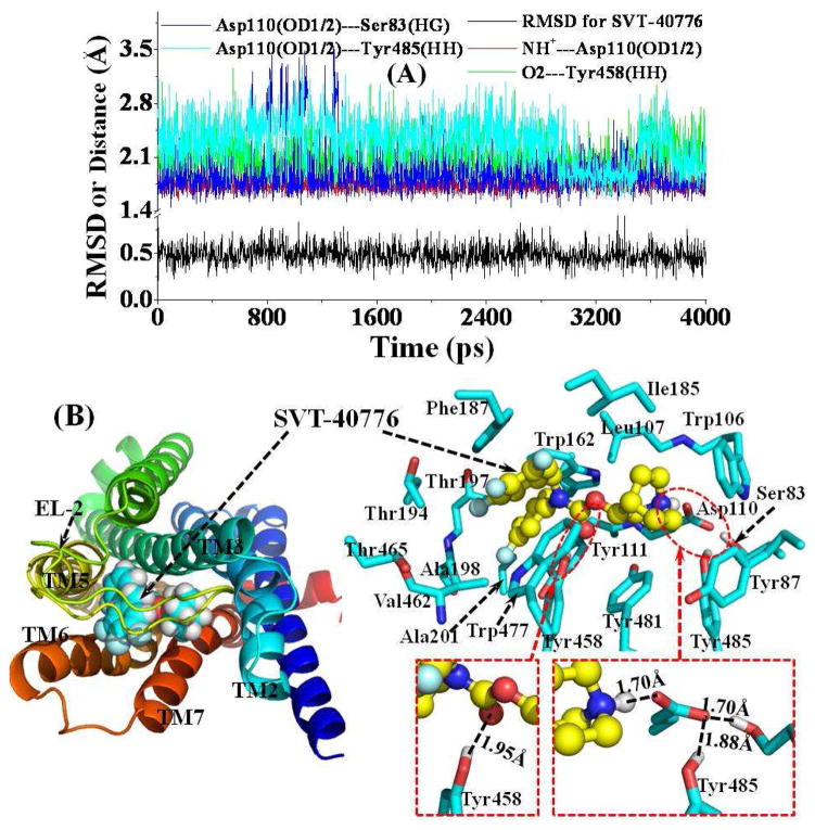
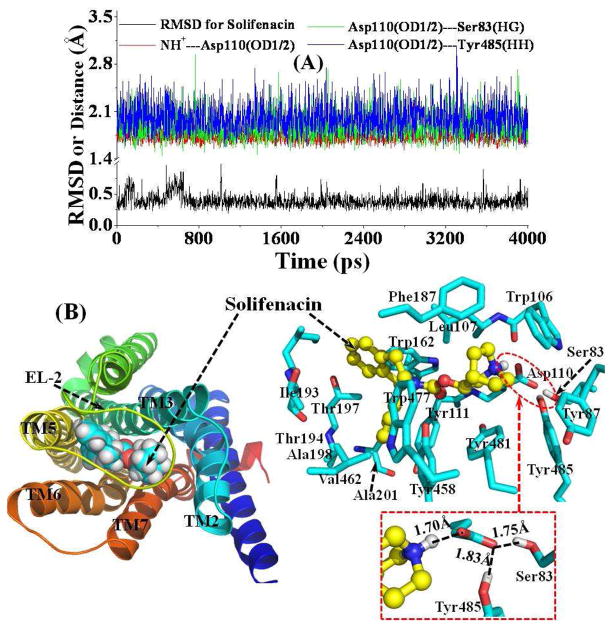
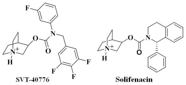
By performing homology modeling, molecular docking, and molecular dynamics (MD) simulations, we have developed three-dimensional (3D) structural models of the M5 muscarinic acetylcholine receptor (mAChR) and two complexes for M5 mAChR binding with antagonists SVT-40776 and solifenacin in the environment of lipid bilayer and solvent water. According to the simulated results, each of the antagonists is oriented horizontally in the binding pocket formed by transmembrane helices 2, 3, and 5-7. The cationic headgroup of each of the antagonists interacts with a negatively charged residue, Asp110, through electrostatic and hydrogen-bonding interactions. The simulated results also reveal some significant difference between the binding modes of SVT-40776 and solifenacin. In particular, SVT-40776 is persistently hydrogen bonded with the side chain of residue Tyr458, whereas solifenacin cannot form a similar hydrogen bond with residues around its carbonyl group. Such significant difference in the binding structures is consistent with the fact that SVT-40776 has a much higher binding affinity (K(d) = 0.4 nM) to M5 mAChR than that of solifenacin (K(d) = 31 nM) with the same reeptor. The calculated binding free energy change (-2.3 ± 0.3 kcal/mol) from solifenacin to SVT-40776 is in good agreement with the experimentally derived binding free energy change (-2.58 kcal/mol), suggesting that our modeled M5 mAChR structure and its complexes with the antagonists are reliable. The new structural insights obtained from this computational study are expected to stimulate further biochemical and pharmacological studies on the detailed structures of M5 and other subtypes of mAChRs.
Figures
Similar articles
-
Novel muscarinic acetylcholine receptor hybrid ligands embedding quinuclidine and 1,4-dioxane fragments.Eur J Med Chem. 2017 Sep 8;137:327-337. doi: 10.1016/j.ejmech.2017.06.004. Epub 2017 Jun 3. Eur J Med Chem. 2017. PMID: 28609709
-
Discovery, synthesis and characterization of a highly muscarinic acetylcholine receptor (mAChR)-selective M5-orthosteric antagonist, VU0488130 (ML381): a novel molecular probe.ChemMedChem. 2014 Aug;9(8):1677-82. doi: 10.1002/cmdc.201402051. Epub 2014 Apr 1. ChemMedChem. 2014. PMID: 24692176 Free PMC article.
-
How dopamine transporter interacts with dopamine: insights from molecular modeling and simulation.Biophys J. 2007 Nov 15;93(10):3627-39. doi: 10.1529/biophysj.107.110924. Epub 2007 Aug 17. Biophys J. 2007. PMID: 17704152 Free PMC article.
-
Understanding G Protein Selectivity of Muscarinic Acetylcholine Receptors Using Computational Methods.Int J Mol Sci. 2019 Oct 24;20(21):5290. doi: 10.3390/ijms20215290. Int J Mol Sci. 2019. PMID: 31653051 Free PMC article.
-
The Muscarinic Acetylcholine Receptor M5: Therapeutic Implications and Allosteric Modulation.ACS Chem Neurosci. 2019 Mar 20;10(3):1025-1034. doi: 10.1021/acschemneuro.8b00481. Epub 2018 Oct 17. ACS Chem Neurosci. 2019. PMID: 30280567 Review.
Cited by
-
Modeling and Re-Engineering of Azotobacter vinelandii Alginate Lyase to Enhance Its Catalytic Efficiency for Accelerating Biofilm Degradation.PLoS One. 2016 Jun 2;11(6):e0156197. doi: 10.1371/journal.pone.0156197. eCollection 2016. PLoS One. 2016. PMID: 27253324 Free PMC article.
-
Metabolites of Vinca Alkaloid Vinblastine: Tubulin Binding and Activation of Nausea-Associated Receptors.ACS Omega. 2019 Jun 4;4(6):9784-9799. doi: 10.1021/acsomega.9b00652. eCollection 2019 Jun 30. ACS Omega. 2019. PMID: 31460070 Free PMC article.
-
Mathematical and computational modeling in biology at multiple scales.Theor Biol Med Model. 2014 Dec 27;11:52. doi: 10.1186/1742-4682-11-52. Theor Biol Med Model. 2014. PMID: 25542608 Free PMC article. Review.
-
Structural modifications to tetrahydropyridine-3-carboxylate esters en route to the discovery of M5-preferring muscarinic receptor orthosteric antagonists.J Med Chem. 2013 Feb 28;56(4):1693-703. doi: 10.1021/jm301774u. Epub 2013 Feb 18. J Med Chem. 2013. PMID: 23379472 Free PMC article.
References
-
- Bonner TI, Young AC, Brann MR, Buckley NJ. Cloning and expression of the human and rat m5 muscarinic acetylcholine receptor genes. Neuron. 1988;1:403–410. - PubMed
-
- Eglen RM, Choppin A, Dillon MP, Hegde S. Muscarinic receptor ligands and their therapeutic potential. Curr Opin Chem Biol. 1999;3:426–432. - PubMed
-
- Bymaster FP, McKinzie DL, Felder CC, Wess J. Use of M1-M5 muscarinic receptor knockout mice as novel tools to delineate the physiological roles of the muscarinic cholinergic system. Neurochem Res. 2003;28:437–442. - PubMed
-
- Jonkam C, Zhu Y, Jacob S, Rehberg S, Kraft E, Hamahata A, Nakano Y, Traber LD, Herndon DN, Traber DL, Hawkins Hk, Enkhbaatar P, Cox RA. Muscarinic receptor antagonist therapy improve acute pulmonary dysfunction after smoke inhalation injury in sheep. Crit Care Med. 2010;38:2339–2344. - PubMed
-
- Yeomans J, Forster G, Blaha C. M5 muscarinic receptors are needed for slow activation of dopamine neurons and for rewarding brain stimulation. Life Sci. 2001;68:2449–2456. - PubMed
Publication types
MeSH terms
Substances
Grants and funding
LinkOut - more resources
Full Text Sources
