Review
. 2012 Jan;139(2):231-43.
doi: 10.1242/dev.067082.

Palatogenesis: morphogenetic and molecular mechanisms of secondary palate development

Affiliations
Review

Palatogenesis: morphogenetic and molecular mechanisms of secondary palate development

Jeffrey O Bush et al. Development. 2012 Jan.

Erratum in

  • Development. 2012 Feb;139(4):828

Abstract

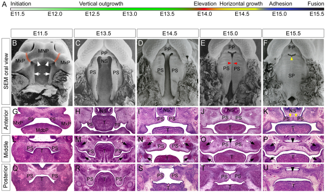
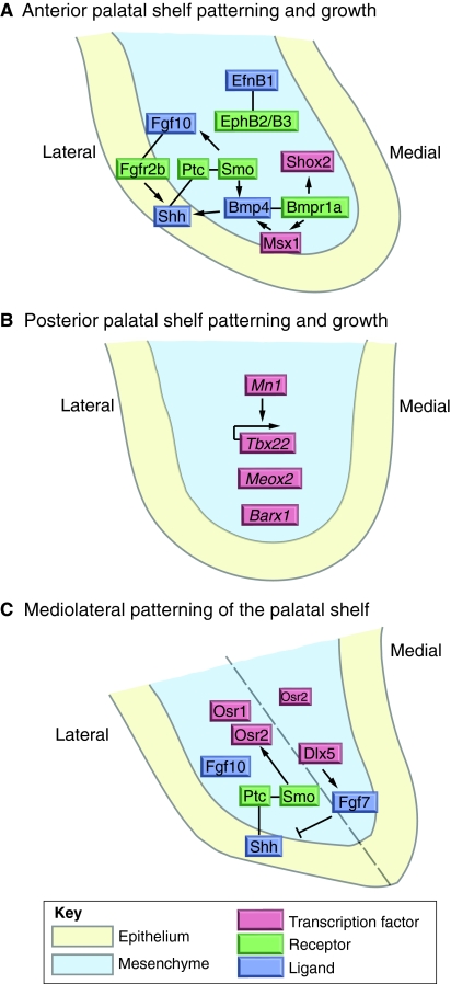
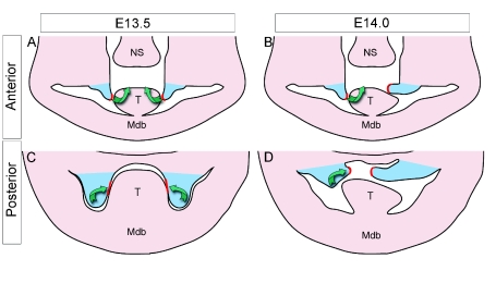
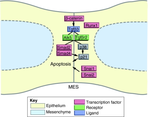
Mammalian palatogenesis is a highly regulated morphogenetic process during which the embryonic primary and secondary palatal shelves develop as outgrowths from the medial nasal and maxillary prominences, respectively, remodel and fuse to form the intact roof of the oral cavity. The complexity of control of palatogenesis is reflected by the common occurrence of cleft palate in humans. Although the embryology of the palate has long been studied, the past decade has brought substantial new knowledge of the genetic control of secondary palate development. Here, we review major advances in the understanding of the morphogenetic and molecular mechanisms controlling palatal shelf growth, elevation, adhesion and fusion, and palatal bone formation.


Figures

Fig. 1.
Fig. 1.
Palatogenesis in the mouse. (A) Timecourse of palate development in mice. (B-F) Scanning electron micrographs showing oral views of the secondary palate at representative developmental stages [reprinted from Kaufman (Kaufman, 1992) with permission]. Orange lines mark sites of fusion between the medial nasal processes and maxillary processes, white arrowheads point to initial outgrowths of the primary palate, white arrows point to the initial outgrowth of the secondary palatal shelves, red arrowheads mark the initial site of palatal adhesion and fusion, and the yellow arrowhead points to the gap between the primary and secondary palates that will disappear following fusion between these tissues. (G-U) Representative histological frontal sections from anterior (G-K), middle (L-P), and posterior (Q-U) regions of the developing palate at each indicated stage. The middle palate region is flanked by the developing upper molar tooth germs (black arrows in M-P) and corresponds to the palatine region of the future hard palate. The posterior palate region corresponds to the future soft palate. At E11.5 (G,L,Q), the palatal shelf outgrowths arise from the oral surface of the maxillary processes. At E13.5 (H,M,R), the palatal shelves exhibit distinct shapes along the AP axis. By E14.5 (I,N,S), the palatal shelves have elevated to the horizontal position. At ∼E15.0 (J,O,T), the palatal shelves make contact at the midline and initiate fusion by formation of the midline epithelial seam (MES) in the mid-anterior region (arrowhead in O). By E15.5 (K,P,U), palatal shelf fusion is evident in the middle and posterior regions, with complete removal of the MES (black arrowheads in P,U). Remnants of the MES can still be seen in the anterior region (K) at this stage and the palatal shelves also fuse superiorly with the nasal septum. Magnification is not equivalent between stages. MdbP, mandibular process; MNP, medial nasal process; MxP, maxillary process; NS, nasal septum; PP, primary palate; PS, palatal shelf; SP, secondary palate; T, tongue.
Fig. 2.
Fig. 2.
Molecular control of palatal shelf growth and patterning. (A) Signaling interactions controlling anterior palatal growth. Sonic hedgehog (Shh) is expressed in the oral epithelium and binds to its receptor patched 1 (Ptc) in the underlying mesenchyme to permit smoothened (Smo) activation of palatal cell proliferation. Fibroblast growth factor 10 (Fgf10) is expressed in the palatal mesenchyme and binds to its receptor Fgfr2b in the palatal epithelium to regulate cell proliferation and survival. Fgf10 and Shh signaling maintain expression of each other to drive palatal outgrowth. Bone morphogenetic protein (Bmp) signaling through the Bmpr1a receptor in the mesenchyme regulates palate growth and expression of Msx1 and Shox2. Bmp signaling is also involved in maintaining Shh expression in the palatal epithelium. Anterior palatal outgrowth is controlled additionally by ephrin B1 (EfnB1) signaling through its receptors EphB2 and EphB3. (B) Genes involved in development of the posterior palate. Mn1, Tbx22, Meox2 and Barx1, which are expressed specifically in the posterior part of the palatal mesenchyme, regulate posterior palatal outgrowth, with Tbx22 acting downstream of Mn1. (C) Pathways responsible for mediolateral patterning of the palatal shelves during vertical outgrowth. Osr1 expression is restricted to the lateral mesenchyme whereas Osr2 is expressed at high levels within the lateral part of the mesenchyme but also in the medial mesenchyme. The expression of both Osr1 and Osr2 is dependent on Shh signaling. An additional pathway involving Dlx5-Fgf7 signaling, which is able to repress Shh signaling, also controls outgrowth along the mediolateral axis. Although not marked in A and B, the mediolateral patterning pathways function throughout the AP axis of the developing palatal shelves; in C we have illustrated the AP axis (dashed line) in the middle palate. Illustrations represent the palatal shelves at specified positions along the AP axis at E13.5. Arrows represent inductive relationships, solid lines represent direct physical interaction, and blunt arrows indicate inhibition.
Fig. 3.
Fig. 3.
Distinct mechanisms for palatal shelf elevation. Palatal shelf elevation at progressive stages, based on drawings from Walker and Fraser (Walker and Fraser, 1956). (A) At late E13, during the beginning of the palatal elevation stage, the anterior palatal shelves (blue) reorient by a ‘flip-up’ mechanism. (B) At ∼E14.0, or mid-elevation, one palatal shelf has just completed elevation, while the other is still vertical. In this flip-up mechanism, the palatal shelves reorient such that the initially distally localized medial edge epithelium (MEE) (red) now becomes the medially located MES epithelium. (C) Initiation of horizontal outgrowth occurs from the medial wall of the vertically oriented palatal shelves in the mid-posterior region. (D) By E14.0, one palatal shelf has completed elevation, while the other is in the process of remodeling. In the posterior palate, the MEE (red) is initially localized to the medial surface of the vertical palatal shelves (Jin et al., 2010) and gives rise to the MES by the remodeling of the underlying mesenchyme. Green arrows depict distinct morphogenetic processes of palatal shelf elevation in the anterior and posterior regions; the anterior palatal shelves elevate in a flip-up process, whereas the posterior palatal shelves undergo remodeling and reorientation through horizontal outgrowth from the medial wall. Mdb, mandible; NS, nasal septum; T, tongue.
Fig. 4.
Fig. 4.
Molecular control of palatal fusion. Tgfβ signaling plays a crucial role in palatal fusion, acting via the Alk5 and Tgfβr2 receptors to activate Smad2/Smad4 and the p38 MAPK pathways, which together regulate p21 expression in the MES. These, in parallel with the transcription factors Snai1 and Snai2, promote MES apoptosis and disintegration. Runx1, which is expressed in the MEE, is required for anterior palatal fusion. Arrows represent inductive relationships and solid lines indicate biochemical activation.

References

    1. Alappat S. R., Zhang Z., Suzuki K., Zhang X., Liu H., Jiang R., Yamada G., Chen Y. (2005). The cellular and molecular etiology of the cleft secondary palate in Fgf10 mutant mice. Dev. Biol. 277, 102-113 - PubMed
    1. Andl T., Ahn K., Kairo A., Chu E. Y., Wine-Lee L., Reddy S. T., Croft N. J., Cebra-Thomas J. A., Metzger D., Chambon P., et al. (2004). Epithelial Bmpr1a regulates differentiation and proliferation in postnatal hair follicles and is essential for tooth development. Development 131, 2257-2268 - PubMed
    1. Baek J. A., Lan Y., Liu H., Maltby K. M., Mishina Y., Jiang R. (2011). Bmpr1a signaling plays critical roles in palatal shelf growth and palatal bone formation. Dev. Biol. 350, 520-531 - PMC - PubMed
    1. Barrow J. R., Stadler H. S., Capecchi M. R. (2000). Roles of Hoxa1 and Hoxa2 in patterning the early hindbrain of the mouse. Development 127, 933-944 - PubMed
    1. Bi W., Huang W., Whitworth D. J., Deng J. M., Zhang Z., Behringer R. R., de Crombrugghe B. (2001). Haploinsufficiency of Sox9 results in defective cartilage primordia and premature skeletal mineralization. Proc. Natl. Acad. Sci. USA 98, 6698-6703 - PMC - PubMed

Publication types

Substances