. 2012 Apr;20(4):749-58.
doi: 10.1038/mt.2011.276. Epub 2011 Dec 20.

The oncolytic poxvirus JX-594 selectively replicates in and destroys cancer cells driven by genetic pathways commonly activated in cancers

Affiliations

The oncolytic poxvirus JX-594 selectively replicates in and destroys cancer cells driven by genetic pathways commonly activated in cancers

Kelley A Parato et al. Mol Ther. 2012 Apr.

Abstract

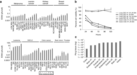
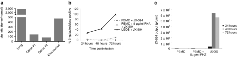
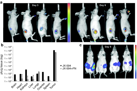
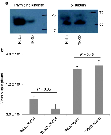
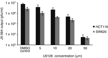
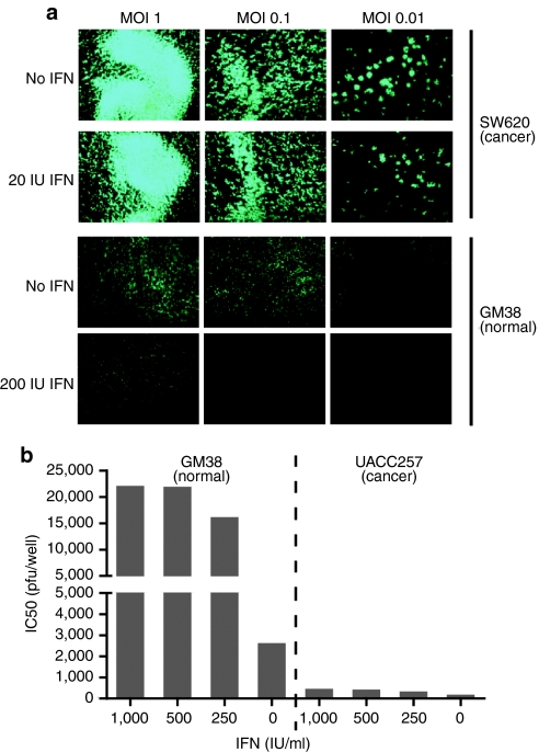
Oncolytic viruses are generally designed to be cancer selective on the basis of a single genetic mutation. JX-594 is a thymidine kinase (TK) gene-inactivated oncolytic vaccinia virus expressing granulocyte-macrophage colony-stimulating factor (GM-CSF) and lac-Z transgenes that is designed to destroy cancer cells through replication-dependent cell lysis and stimulation of antitumoral immunity. JX-594 has demonstrated a favorable safety profile and reproducible tumor necrosis in a variety of solid cancer types in clinical trials. However, the mechanism(s) responsible for its cancer-selectivity have not yet been well described. We analyzed the replication of JX-594 in three model systems: primary normal and cancer cells, surgical explants, and murine tumor models. JX-594 replication, transgene expression, and cytopathic effects were highly cancer-selective, and broad spectrum activity was demonstrated. JX-594 cancer-selectivity was multi-mechanistic; replication was activated by epidermal growth factor receptor (EGFR)/Ras pathway signaling, cellular TK levels, and cancer cell resistance to type-I interferons (IFNs). These findings confirm a large therapeutic index for JX-594 that is driven by common genetic abnormalities in human solid tumors. This appears to be the first description of multiple selectivity mechanisms, both inherent and engineered, for an oncolytic virus. These findings have implications for oncolytic viruses in general, and suggest that their cancer targeting is a complex and multifactorial process.


Figures

Figure 1
Figure 1
JX-594 replicates and induces cytolysis in a broad spectrum of cancer cell types, including cell lines, tumor initiating cells, and primary human tumor tissue. (a) JX-594 ED50 (pfu/cell) on human cell lines in the NCI cell line panel upon JX-594 infection at multiple multiplicities of infection (MOI) 72 hours postinfection (n = 3). Ras mutational status for colorectal cancer cell lines is indicated. Error bars = SD. (b) Percent viability (compared to untreated control) over time of three tumor initiating cell lines (lung or colon) following infection with JX-594 or UV-inactivated JX-594 control at an MOI of 3 (n =3). Error bars = SD. (c) Human tumor biopsy material was sliced into 5 × 5 × 5 mm segments and infected with 5 × 10⁶ pfu JX-594 suspended in 100 µl α-MEM serum-free medium ex vivo. Samples were homogenized and infectious virus quantitated 48 hours postinfection. MEM, minimum essential medium; NCI, National Cancer Institute; NSCLC, non-small-cell lung cancer; UV, ultraviolet.
Figure 2
Figure 2
JX-594 preferentially infects tumor tissue, while sparing normal tissue. (a) Human tumor biopsy material, and when possible accompanying adjacent normal organ tissue samples, were sliced into 5 × 5 × 5 mm segments and infected with 5 × 10⁶ pfu JX-594 or JX-594-GFP+/β-gal ex vivo. When possible, paired normal and tumor tissue samples wherein the tumor displayed virus-driven fluorescent reporter expression, were homogenized and titered to quantify infectious virus recovered from the specimen 48 hours postinfection. The ratio of JX-594-GFP+/β-gal in tumor tissue versus the corresponding normal tissue is plotted. (b) Donor PBMC from a healthy volunteer were cultured for 24 hours in the presence or absence of PHA (5 µg/ml) before infection with JX-594. PBMC or U2OS cell cultures were then inoculated with JX-594 at an MOI = 1 pfu/cell and after 24, 48, or 72 hours. β-gal activity was detected by flow cytometry using ImaGene Green LacZ detection kit (n =1). (c) JX-594 output from duplicate culture/infection conditions as measured by plaque assay (n = 1). β-gal, β-galactosidase; GFP, green fluorescent protein; MOI, multiplicity of infection; PBMC, peripheral blood mononuclear cells; PHA, phytohemagglutinin.
Figure 3
Figure 3
In vivo biodistribution of JX-594-luc+/β-gal infection and replication following intravenous administration. (a) Athymic nude mice with established subcutaneous SW620 human colon carcinoma tumors were treated intravenously via the tail vein with 107 pfu JX-594-luc+/β-gal. Three and 6 days postinfection, luminescence was assessed emanating from the tumor and normal tissue with IVIS system. (b) Athymic nude mice bearing subcutaneous SW620 tumors treated with intravenous JX-594-luc+/β-gal at a dose of 1 × 10⁷ pfu in a, as well as mice treated with JX-594-luc+/β-gal and universal IFN-α were sacrificed 7 days following JX-594-luc+/β-gal treatment. Organs were collected and the presence of virus was quantified by plaque assay in U2OS cells (n = 6 per group). Error bars = SD. (c) Female transgenic FVB/N mice with autochthonous ovarian tumors were treated with 1 × 10⁸ pfu JX-594-luc+/β-gal intraperitoneally, and 5 days later luminescence was assessed with a Xenogen 200 Series IVIS system. β-gal, β-galactosidase; IFN, interferon; IVIS, in vivo imaging system; luc, firefly luciferase; MOI, multiplicity of infection; shRNA, short hairpin RNA; TK, thymidine kinase; TKKD, thymidine kinase knockdown.
Figure 4
Figure 4
JX-594 replication is attenuated in cancer cells depleted of cellular thymidine kinase (TK). (a) Western blotting confirming knockdown of endogenous TK in HeLa cells performed by retroviral transduction using shRNA against TK, or a control shRNA. (b) JX-594 and wild-type Wyeth control replication in HeLa cells or TK-knockdown clones, following infection at MOI 0.1 for 48 hours before performing plaque assays on infected material (n =3). Error bars = SD. MOI, multiplicity of infection; shRNA, short hairpin RNA.
Figure 5
Figure 5
JX-594-GFP+/β-gal replication is attenuated in the presence of an Erk inhibitor. Two human colon carcinoma cell lines (SW620 and HCT116) were infected with JX-594-GFP+/β-gal at an MOI of 0.1 for 50 hours in the presence or absence of the Erk inhibitor U0126. JX-594-GFP+/β-gal output from triplicate culture/infection conditions was measured by plaque assay (n = 3). Error bars = SD. β-gal, β-galactosidase; DMSO, dimethyl sulfoxide; Erk, extracellular signal-regulated kinase; GFP, green fluorescent protein.
Figure 6
Figure 6
JX-594-GFP+/β-gal replication is attenuated in the presence of interferon on normal cells, but not on cancer cells. (a) SW620 human colon carcinoma cells (interferon-resistant) and GM38 normal human fibroblasts (interferon-sensitive) were inoculated with JX-594-GFP+/β-gal (MOI 0.001–1) in the presence or absence of 200 IU/ml exogenous IFN-α and transgene expression (GFP) was monitored 48 hours postinfection using a Zeiss Axiovision microscope and Axiovision acquisition and image storage software. (b) GM38 normal human fibroblasts and UACC257 human melanoma cells (interferon-resistant) were seeded (3 × 10⁴ cells per well) in the presence or absence of varying concentrations of IFN-α and infected with JX-594-GFP+/β-gal at various MOIs, and 72 hours later cell viability was assessed by CellTiter Blue assay (n = 4). β-gal, β-galactosidase; GFP, green fluorescent protein; IFN, interferon; MOI, multiplicity of infection.

References

    1. Parato KA, Lichty BD., and, Bell JC. Diplomatic immunity: turning a foe into an ally. Curr Opin Mol Ther. 2009;11:13–21. - PubMed
    1. Cattaneo R, Miest T, Shashkova EV., and, Barry MA. Reprogrammed viruses as cancer therapeutics: targeted, armed and shielded. Nat Rev Microbiol. 2008;6:529–540. - PMC - PubMed
    1. Bell JC, Lichty B., and, Stojdl D. Getting oncolytic virus therapies off the ground. Cancer Cell. 2003;4:7–11. - PubMed
    1. Liu TC, Hwang TH, Bell JC., and, Kirn DH. Development of targeted oncolytic virotherapeutics through translational research. Expert Opin Biol Ther. 2008;8:1381–1391. - PubMed
    1. Liu TC, Galanis E., and, Kirn D. Clinical trial results with oncolytic virotherapy: a century of promise, a decade of progress. Nat Clin Pract Oncol. 2007;4:101–117. - PubMed

Publication types

MeSH terms